 Movie
Movie Controller

[English] 日本語
 Yorodumi
- PDB-4oj6: Crystal Structure of a Putative Tailspike Protein (TSP1, orf210) ... -
+ Open data

- Basic information
| Entry | Database: PDB / ID: 4oj6 | ||||||
|---|---|---|---|---|---|---|---|
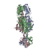
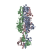
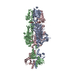
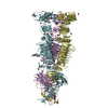
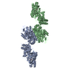
| Title | Crystal Structure of a Putative Tailspike Protein (TSP1, orf210) from Escherichia coli O157:H7 Bacteriohage CBA120; Se-Met Protein | ||||||
 Components Components | Tailspike protein | ||||||
 Keywords Keywords |  VIRAL PROTEIN / parallel beta helix / putative endo-glycosidase / bacterial polysaccharide / phage baseplate / phage tail VIRAL PROTEIN / parallel beta helix / putative endo-glycosidase / bacterial polysaccharide / phage baseplate / phage tail | ||||||
| Function / homology |  Function and homology information Function and homology informationbiological process involved in interaction with host / viral life cycle / virion componentSimilarity search - Function | ||||||
| Biological species |  Escherichia phage Cba120 (virus) Escherichia phage Cba120 (virus) | ||||||
| Method | X-RAY DIFFRACTION / SYNCHROTRON / MAD / Resolution: 1.8 Å | ||||||
 Authors Authors | Chen, C. / Herzberg, O. | ||||||
 Citation Citation | Journal: Plos One / Year: 2014 Title: Crystal structure of ORF210 from E. coli O157:H1 phage CBA120 (TSP1), a putative tailspike protein. Authors: Chen, C. / Bales, P. / Greenfield, J. / Heselpoth, R.D. / Nelson, D.C. / Herzberg, O. | ||||||
| History |
|
- Structure visualization
| Structure viewer | Molecule: MolmilJmol/JSmol |
|---|
- Downloads & links
-Download
| PDBx/mmCIF format | 4oj6.cif.gz | 828.4 KB | Display | PDBx/mmCIF format |
|---|---|---|---|---|
| PDB format | pdb4oj6.ent.gz | 707.8 KB | Display | PDB format |
| PDBx/mmJSON format | 4oj6.json.gz | Tree view | PDBx/mmJSON format | |
| Others |  Other downloads Other downloads |
-Validation report
| Arichive directory | https://data.pdbj.org/pub/pdb/validation_reports/oj/4oj6ftp://data.pdbj.org/pub/pdb/validation_reports/oj/4oj6 | HTTPS FTP |
|---|
-Related structure data


-Links
 PDBj
PDBj- Assembly
| Deposited unit | 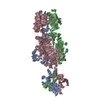
| ||||||||
|---|---|---|---|---|---|---|---|---|---|
| 1 |
| ||||||||
| Unit cell |
|
-Components
| #1: Protein | Mass: 83417.852 Da / Num. of mol.: 3 Source method: isolated from a genetically manipulated source Source: (gene. exp.) Escherichia phage Cba120 (virus) / Gene: orf210 / Plasmid: pBAD24 / Production host:  Escherichia coli (E. coli) / Strain (production host): BL21 / References: UniProt: G3M189 Escherichia coli (E. coli) / Strain (production host): BL21 / References: UniProt: G3M189#2: Chemical | ChemComp-ZN / |   Mass: 65.409 Da / Num. of mol.: 1 / Source method: obtained synthetically / Formula: Zn Mass: 65.409 Da / Num. of mol.: 1 / Source method: obtained synthetically / Formula: Zn#3: Water | ChemComp-HOH / | Water Mass: 18.015 Da / Num. of mol.: 2362 / Source method: isolated from a natural source / Formula: H2O Mass: 18.015 Da / Num. of mol.: 2362 / Source method: isolated from a natural source / Formula: H2O |
|---|
-Experimental details
-Experiment
| Experiment | Method: X-RAY DIFFRACTION / Number of used crystals: 1 |
|---|
- Sample preparation
| Crystal | Density Matthews: 3.23 Å3/Da / Density % sol: 61.95 % |
|---|---|
| Crystal grow | Temperature: 298 K / Method: vapor diffusion, hanging drop / pH: 7 Details: 0.1 M Tris pH 7.0-7.6, 20% PEG-1000, VAPOR DIFFUSION, HANGING DROP, temperature 298K |
-Data collection
| Diffraction | Mean temperature: 100 K | |||||||||||||||
|---|---|---|---|---|---|---|---|---|---|---|---|---|---|---|---|---|
| Diffraction source | Source: SYNCHROTRON / Site: APS  / Beamline: 23-ID-B / Wavelength: 0.97945, 0.97961, 0.94945, 1.03320 / Beamline: 23-ID-B / Wavelength: 0.97945, 0.97961, 0.94945, 1.03320 | |||||||||||||||
| Detector | Type: MARMOSAIC 300 mm CCD / Detector: CCD / Date: Nov 10, 2012 / Details: mirrors | |||||||||||||||
| Radiation | Monochromator: Si 1 1 1 / Protocol: MAD / Monochromatic (M) / Laue (L): M / Scattering type: x-ray | |||||||||||||||
| Radiation wavelength |
| |||||||||||||||
| Reflection | Resolution: 1.8→90 Å / Num. all: 298429 / Num. obs: 297461 / % possible obs: 99.6 % / Observed criterion σ(F): 2 / Observed criterion σ(I): 2 / Redundancy: 6.5 % / Rmerge(I) obs: 0.087 / Net I/σ(I): 13.3 | |||||||||||||||
| Reflection shell | Resolution: 1.8→1.85 Å / Redundancy: 4.2 % / Rmerge(I) obs: 0.5 / Mean I/σ(I) obs: 2.1 / Num. unique all: 21821 / Rsym value: 0.5 / % possible all: 99.7 |
- Processing
| Software |
| |||||||||||||||||||||||||||||||||||||||||||||||||||||||||||||||||||||||||||||||||||||||||||||||||||||||||||||||||||||||||||||||||||||||||||||||||||||||||||||||||||||||||||||||||||||||||||||||||||||||||||||||||||||||||
|---|---|---|---|---|---|---|---|---|---|---|---|---|---|---|---|---|---|---|---|---|---|---|---|---|---|---|---|---|---|---|---|---|---|---|---|---|---|---|---|---|---|---|---|---|---|---|---|---|---|---|---|---|---|---|---|---|---|---|---|---|---|---|---|---|---|---|---|---|---|---|---|---|---|---|---|---|---|---|---|---|---|---|---|---|---|---|---|---|---|---|---|---|---|---|---|---|---|---|---|---|---|---|---|---|---|---|---|---|---|---|---|---|---|---|---|---|---|---|---|---|---|---|---|---|---|---|---|---|---|---|---|---|---|---|---|---|---|---|---|---|---|---|---|---|---|---|---|---|---|---|---|---|---|---|---|---|---|---|---|---|---|---|---|---|---|---|---|---|---|---|---|---|---|---|---|---|---|---|---|---|---|---|---|---|---|---|---|---|---|---|---|---|---|---|---|---|---|---|---|---|---|---|---|---|---|---|---|---|---|---|---|---|---|---|---|---|---|---|
| Refinement | Method to determine structure: MAD / Resolution: 1.8→19.851 Å / SU ML: 0.24 / Cross valid method: THROUGHOUT / σ(F): 1.46 / Phase error: 22.18 / Stereochemistry target values: Engh & Huber
| |||||||||||||||||||||||||||||||||||||||||||||||||||||||||||||||||||||||||||||||||||||||||||||||||||||||||||||||||||||||||||||||||||||||||||||||||||||||||||||||||||||||||||||||||||||||||||||||||||||||||||||||||||||||||
| Solvent computation | Shrinkage radii: 0.9 Å / VDW probe radii: 1.11 Å / Solvent model: FLAT BULK SOLVENT MODEL | |||||||||||||||||||||||||||||||||||||||||||||||||||||||||||||||||||||||||||||||||||||||||||||||||||||||||||||||||||||||||||||||||||||||||||||||||||||||||||||||||||||||||||||||||||||||||||||||||||||||||||||||||||||||||
| Refinement step | Cycle: LAST / Resolution: 1.8→19.851 Å
| |||||||||||||||||||||||||||||||||||||||||||||||||||||||||||||||||||||||||||||||||||||||||||||||||||||||||||||||||||||||||||||||||||||||||||||||||||||||||||||||||||||||||||||||||||||||||||||||||||||||||||||||||||||||||
| Refine LS restraints |
| |||||||||||||||||||||||||||||||||||||||||||||||||||||||||||||||||||||||||||||||||||||||||||||||||||||||||||||||||||||||||||||||||||||||||||||||||||||||||||||||||||||||||||||||||||||||||||||||||||||||||||||||||||||||||
| LS refinement shell |
|
