 Movie
Movie Controller

[English] 日本語
 Yorodumi
- PDB-4oit: Structure, interactions and evolutionary implications of a domain... -
+ Open data

- Basic information
| Entry | Database: PDB / ID: 4oit | ||||||
|---|---|---|---|---|---|---|---|
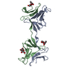
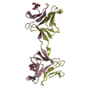
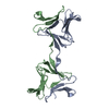
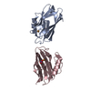
| Title | Structure, interactions and evolutionary implications of a domain-swapped lectin dimer from Mycobacterium smegmatis | ||||||
 Components Components | LysM domain protein | ||||||
 Keywords Keywords | SUGAR BINDING PROTEIN / Beta-prism II fold / bacterial lectin / protein-carbohydrate interactions / Beta-Prism II / Carbohydrate binding / Carbohydrate/Sugar | ||||||
| Function / homology |  Function and homology informationAgglutinin, subunit A / Bulb-type lectin domain / Bulb-type lectin domain / Bulb-type lectin domain superfamily / Bulb-type lectin domain profile. / Bulb-type mannose-specific lectin / Orthogonal Prism / Lysin motif / LysM domain superfamily / LysM domain ...Agglutinin, subunit A / Bulb-type lectin domain / Bulb-type lectin domain / Bulb-type lectin domain superfamily / Bulb-type lectin domain profile. / Bulb-type mannose-specific lectin / Orthogonal Prism / Lysin motif / LysM domain superfamily / LysM domain / LysM domain profile. / LysM domain / Mainly Beta Function and homology informationAgglutinin, subunit A / Bulb-type lectin domain / Bulb-type lectin domain / Bulb-type lectin domain superfamily / Bulb-type lectin domain profile. / Bulb-type mannose-specific lectin / Orthogonal Prism / Lysin motif / LysM domain superfamily / LysM domain ...Agglutinin, subunit A / Bulb-type lectin domain / Bulb-type lectin domain / Bulb-type lectin domain superfamily / Bulb-type lectin domain profile. / Bulb-type mannose-specific lectin / Orthogonal Prism / Lysin motif / LysM domain superfamily / LysM domain / LysM domain profile. / LysM domain / Mainly BetaSimilarity search - Domain/homology | ||||||
| Biological species |  Mycobacterium smegmatis (bacteria) Mycobacterium smegmatis (bacteria) | ||||||
| Method | X-RAY DIFFRACTION / MOLECULAR REPLACEMENT / Resolution: 2.24 Å | ||||||
 Authors Authors | Patra, D. / Mishra, P. / Surolia, A. / Vijayan, M. | ||||||
 Citation Citation | Journal: Glycobiology / Year: 2014 Title: Structure, interactions and evolutionary implications of a domain-swapped lectin dimer from Mycobacterium smegmatis. Authors: Patra, D. / Mishra, P. / Surolia, A. / Vijayan, M. #1: Journal: Acta Crystallogr.,Sect.F / Year: 2011 Title: Cloning, expression, purification, crystallization and preliminary X-ray studies of the mannose-binding lectin domain of MSMEG_3662 from Mycobacterium smegmatis Authors: Patra, D. / Sharma, A. / Chandran, D. / Vijayan, M. #2: Journal: J.Biosci. / Year: 2007 Title: Multiplicity of carbohydrate-binding sites in beta-prism fold lectins: occurrence and possible evolutionary implications Authors: Sharma, A. / Chandran, D. / Singh, D.D. / Vijayan, M. #3: Journal: Proteins / Year: 2013 Title: Identification of mycobacterial lectins from genomic data Authors: Abhinav, K.V. / Sharma, A. / Vijayan, M. | ||||||
| History |
|
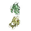
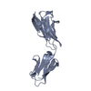
- Structure visualization
| Structure viewer | Molecule: MolmilJmol/JSmol |
|---|
- Downloads & links
-Download
| PDBx/mmCIF format | 4oit.cif.gz | 95.2 KB | Display | PDBx/mmCIF format |
|---|---|---|---|---|
| PDB format | pdb4oit.ent.gz | 74 KB | Display | PDB format |
| PDBx/mmJSON format | 4oit.json.gz | Tree view | PDBx/mmJSON format | |
| Others |  Other downloads Other downloads |
-Validation report
| Arichive directory | https://data.pdbj.org/pub/pdb/validation_reports/oi/4oitftp://data.pdbj.org/pub/pdb/validation_reports/oi/4oit | HTTPS FTP |
|---|
-Related structure data


-Links
 PDBj
PDBj


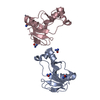
- Assembly
| Deposited unit | 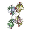
| ||||||||
|---|---|---|---|---|---|---|---|---|---|
| 1 | 
| ||||||||
| 2 | 
| ||||||||
| Unit cell |
|
-Components
| #1: Protein | / Mannose-binding lectin Mass: 12541.867 Da / Num. of mol.: 4 / Fragment: mannose-binding lectin domain, UNP residues 1-105 Source method: isolated from a genetically manipulated source Source: (gene. exp.) Mycobacterium smegmatis (bacteria) / Strain: MC2 155 / Gene: MSMEG_3662 / Plasmid: pET21b / Production host: Escherichia coli (E. coli) / Strain (production host): BL21(DE3) / References: UniProt: A0QYH7#2: Sugar | ChemComp-MAN / Mannose  Type: D-saccharide, alpha linking / Mass: 180.156 Da / Num. of mol.: 10 Type: D-saccharide, alpha linking / Mass: 180.156 Da / Num. of mol.: 10Source method: isolated from a genetically manipulated source Formula: C6H12O6 #3: Sugar | ChemComp-BMA / | Mannose  Type: D-saccharide, beta linking / Mass: 180.156 Da / Num. of mol.: 1 Type: D-saccharide, beta linking / Mass: 180.156 Da / Num. of mol.: 1Source method: isolated from a genetically manipulated source Formula: C6H12O6 #4: Water | ChemComp-HOH / | Water Mass: 18.015 Da / Num. of mol.: 130 / Source method: isolated from a natural source / Formula: H2O Mass: 18.015 Da / Num. of mol.: 130 / Source method: isolated from a natural source / Formula: H2O |
|---|
-Experimental details
-Experiment
| Experiment | Method: X-RAY DIFFRACTION / Number of used crystals: 1 |
|---|
- Sample preparation
| Crystal | Density Matthews: 2.05 Å3/Da / Density % sol: 40.11 % |
|---|---|
| Crystal grow | Temperature: 298 K / Method: vapor diffusion, hanging drop / pH: 7.5 Details: 1.2M tri-sodium citrate, 0.1M Na HEPES, 6% glycerol, 30%(w/v) 1,5 diammino pentane dihydrochloride, 60mM mannose, pH 7.5, VAPOR DIFFUSION, HANGING DROP, temperature 298K |
-Data collection
| Diffraction | Mean temperature: 100 K |
|---|---|
| Diffraction source | Source: ROTATING ANODE / Type: BRUKER AXS MICROSTAR / Wavelength: 1.54179 Å |
| Detector | Type: MAR scanner 345 mm plate / Detector: IMAGE PLATE / Date: Mar 11, 2012 / Details: mirrors |
| Radiation | Monochromator: Mirrors / Protocol: SINGLE WAVELENGTH / Monochromatic (M) / Laue (L): M / Scattering type: x-ray |
| Radiation wavelength | Wavelength: 1.54179 Å / Relative weight: 1 |
| Reflection | Resolution: 2.24→40 Å / Num. all: 47027 / Num. obs: 19379 / % possible obs: 98.7 % / Redundancy: 2.7 % / Rmerge(I) obs: 0.112 / Net I/σ(I): 6.2 |
| Reflection shell | Resolution: 2.24→2.36 Å / Redundancy: 2.3 % / Rmerge(I) obs: 0.38 / Num. unique all: 2766 / % possible all: 97.2 |
- Processing
| Software |
| |||||||||||||||||||||||||||||||||||||||||||||||||||||||||||||||||
|---|---|---|---|---|---|---|---|---|---|---|---|---|---|---|---|---|---|---|---|---|---|---|---|---|---|---|---|---|---|---|---|---|---|---|---|---|---|---|---|---|---|---|---|---|---|---|---|---|---|---|---|---|---|---|---|---|---|---|---|---|---|---|---|---|---|---|
| Refinement | Method to determine structure: MOLECULAR REPLACEMENT Starting model: Native structure Resolution: 2.24→40 Å / Cor.coef. Fo:Fc: 0.915 / Cor.coef. Fo:Fc free: 0.882 / SU B: 7.79 / SU ML: 0.197 / Cross valid method: THROUGHOUT / ESU R: 0.44 / ESU R Free: 0.259 / Stereochemistry target values: MAXIMUM LIKELIHOOD / Details: HYDROGENS HAVE BEEN ADDED IN THE RIDING POSITIONS
| |||||||||||||||||||||||||||||||||||||||||||||||||||||||||||||||||
| Solvent computation | Ion probe radii: 0.8 Å / Shrinkage radii: 0.8 Å / VDW probe radii: 1.4 Å / Solvent model: MASK | |||||||||||||||||||||||||||||||||||||||||||||||||||||||||||||||||
| Displacement parameters | Biso mean: 22.834 Å2
| |||||||||||||||||||||||||||||||||||||||||||||||||||||||||||||||||
| Refinement step | Cycle: LAST / Resolution: 2.24→40 Å
| |||||||||||||||||||||||||||||||||||||||||||||||||||||||||||||||||
| Refine LS restraints |
| |||||||||||||||||||||||||||||||||||||||||||||||||||||||||||||||||
| LS refinement shell | Resolution: 2.24→2.298 Å / Total num. of bins used: 20
|
