- PDB-4oen: Crystal structure of the second substrate binding domain of a put... -
+
Open data
ID or keywords:
Loading...
-
Basic information
Entry
Database: PDB / ID: 4oen
Title
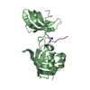
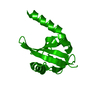
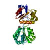
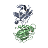
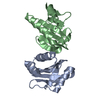
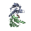
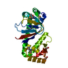
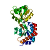
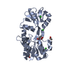
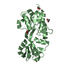
Crystal structure of the second substrate binding domain of a putative amino acid ABC transporter from Streptococcus pneumoniae Canada MDR_19A
Components
Second substrate binding domain of putative amino acid ABC transporter
Keywords
TRANSPORT PROTEIN / CENTER FOR STRUCTURAL GENOMICS OF INFECTIOUS DISEASES (CSGID) / NIAID / NATIONAL INSTITUTE OF ALLERGY AND INFECTIOUS DISEASES / ALPHA AND BETA PROTEIN / PERIPLASMIC BINDING PROTEIN TYPE II FOLD / SUBSTRATE BINDING DOMAIN OF PUTATIVE AMINO ACID ABC TRANSPORTER SYSTEM
Function / homology
Function and homology information
amino acid transport / ligand-gated monoatomic ion channel activity / ATP-binding cassette (ABC) transporter complex Similarity search - Function
ABC transporter membrane protein permease protein ArtM/GltK/GlnP/TcyL/YhdX-like / Amino acid ABC transporter, permease protein, 3-TM domain / ABC transporter type 1, transmembrane domain MetI-like / MetI-like superfamily / Binding-protein-dependent transport system inner membrane component / ABC transporter integral membrane type-1 domain profile. / Bacterial periplasmic substrate-binding proteins / Bacterial extracellular solute-binding proteins, family 3 / Solute-binding protein family 3/N-terminal domain of MltF / Ionotropic glutamate receptor ...ABC transporter membrane protein permease protein ArtM/GltK/GlnP/TcyL/YhdX-like / Amino acid ABC transporter, permease protein, 3-TM domain / ABC transporter type 1, transmembrane domain MetI-like / MetI-like superfamily / Binding-protein-dependent transport system inner membrane component / ABC transporter integral membrane type-1 domain profile. / Bacterial periplasmic substrate-binding proteins / Bacterial extracellular solute-binding proteins, family 3 / Solute-binding protein family 3/N-terminal domain of MltF / Ionotropic glutamate receptor / Eukaryotic homologues of bacterial periplasmic substrate binding proteins. / Periplasmic binding protein-like II / D-Maltodextrin-Binding Protein; domain 2 / 3-Layer(aba) Sandwich / Alpha Beta Similarity search - Domain/homology
In the structure databanks used in Yorodumi, some data are registered as the other names, "COVID-19 virus" and "2019-nCoV". Here are the details of the virus and the list of structure data.
Jan 31, 2019. EMDB accession codes are about to change! (news from PDBe EMDB page)
EMDB accession codes are about to change! (news from PDBe EMDB page)
The allocation of 4 digits for EMDB accession codes will soon come to an end. Whilst these codes will remain in use, new EMDB accession codes will include an additional digit and will expand incrementally as the available range of codes is exhausted. The current 4-digit format prefixed with “EMD-” (i.e. EMD-XXXX) will advance to a 5-digit format (i.e. EMD-XXXXX), and so on. It is currently estimated that the 4-digit codes will be depleted around Spring 2019, at which point the 5-digit format will come into force.
The EM Navigator/Yorodumi systems omit the EMD- prefix.
Related info.:Q: What is EMD? / ID/Accession-code notation in Yorodumi/EM Navigator
Yorodumi is a browser for structure data from EMDB, PDB, SASBDB, etc.
This page is also the successor to EM Navigator detail page, and also detail information page/front-end page for Omokage search.
The word "yorodu" (or yorozu) is an old Japanese word meaning "ten thousand". "mi" (miru) is to see.
Related info.:EMDB / PDB / SASBDB / Comparison of 3 databanks / Yorodumi Search / Aug 31, 2016. New EM Navigator & Yorodumi / Yorodumi Papers / Jmol/JSmol / Function and homology information / Changes in new EM Navigator and Yorodumi
 Movie
Movie Controller

 Yorodumi
 Open data
 Basic information
 Components
 Keywords
 TRANSPORT PROTEIN / CENTER FOR STRUCTURAL GENOMICS OF INFECTIOUS DISEASES (CSGID) / NIAID /
  Function and homology information

 Authors
 Citation
 Structure visualization
 Downloads & links
 Other downloads
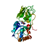
 PDBj
PDBj
 Assembly
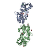

 Mass: 96.063 Da / Num. of mol.: 2 / Source method: obtained synthetically / Formula: SO4
 Mass: 35.453 Da / Num. of mol.: 8 / Source method: obtained synthetically / Formula: Cl
 Mass: 59.044 Da / Num. of mol.: 6 / Source method: obtained synthetically / Formula: C2H3O2
 Mass: 18.015 Da / Num. of mol.: 788 / Source method: isolated from a natural source / Formula: H2O
 Sample preparation
 / Beamline: 21-ID-F / Wavelength: 0.97872 Å
 Processing