 Movie
Movie Controller

[English] 日本語
 Yorodumi
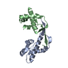
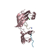
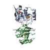
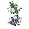
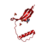
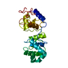
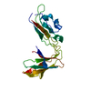
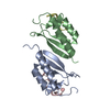
- PDB-4odk: Structure of SlyD from Thermus thermophilus in complex with T1 peptide -
+ Open data

- Basic information
| Entry | Database: PDB / ID: 4odk | ||||||
|---|---|---|---|---|---|---|---|
| Title | Structure of SlyD from Thermus thermophilus in complex with T1 peptide | ||||||
 Components Components |
| ||||||
 Keywords Keywords |  ISOMERASE / CHAPERONE / FKBP domain / IF domain / peptidyl-prolyl isomerase / PPIase ISOMERASE / CHAPERONE / FKBP domain / IF domain / peptidyl-prolyl isomerase / PPIase | ||||||
| Function / homology |  Function and homology information Function and homology informationhyphal tip / ribonuclease T1 activity / ribonuclease T1 / cell septum / peptidylprolyl isomerase / peptidyl-prolyl cis-trans isomerase activity / protein refolding / endonuclease activity / lyase activity / RNA binding / metal ion bindingSimilarity search - Function | ||||||
| Biological species |   Thermus thermophilus (bacteria) Thermus thermophilus (bacteria) Aspergillus oryzae (mold) Aspergillus oryzae (mold) | ||||||
| Method | X-RAY DIFFRACTION / SYNCHROTRON / MOLECULAR REPLACEMENT / Resolution: 1.401 Å | ||||||
 Authors Authors | Quistgaard, E.M. / Low, C. / Nordlund, P. | ||||||
 Citation Citation | Journal: BMC Biol. / Year: 2016 Title: Molecular insights into substrate recognition and catalytic mechanism of the chaperone and FKBP peptidyl-prolyl isomerase SlyD. Authors: Quistgaard, E.M. / Weininger, U. / Ural-Blimke, Y. / Modig, K. / Nordlund, P. / Akke, M. / Low, C. | ||||||
| History |
|
- Structure visualization
| Structure viewer | Molecule: MolmilJmol/JSmol |
|---|
- Downloads & links
-Download
| PDBx/mmCIF format | 4odk.cif.gz | 85.1 KB | Display | PDBx/mmCIF format |
|---|---|---|---|---|
| PDB format | pdb4odk.ent.gz | 68.2 KB | Display | PDB format |
| PDBx/mmJSON format | 4odk.json.gz | Tree view | PDBx/mmJSON format | |
| Others |  Other downloads Other downloads |
-Validation report
| Arichive directory | https://data.pdbj.org/pub/pdb/validation_reports/od/4odkftp://data.pdbj.org/pub/pdb/validation_reports/od/4odk | HTTPS FTP |
|---|
-Related structure data
| Related structure data |  4odlC 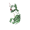 4odmC 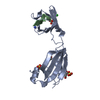 4odnC 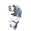 4odoC 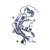 4odpC  4odqC 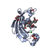 4odrC C: citing same article ( |
|---|---|
| Similar structure data |

-Links
 PDBj
PDBj


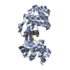
- Assembly
| Deposited unit | 
| ||||||||
|---|---|---|---|---|---|---|---|---|---|
| 1 |
| ||||||||
| Unit cell |
|
-Components
| #1: Protein | Mass: 17400.234 Da / Num. of mol.: 1 Source method: isolated from a genetically manipulated source Source: (gene. exp.) Thermus thermophilus (bacteria) / Gene: TTHA0346 / Production host: Escherichia coli (E. coli) / References: UniProt: Q5SLE7, peptidylprolyl isomerase | ||||
|---|---|---|---|---|---|
| #2: Protein/peptide | Mass: 1728.798 Da / Num. of mol.: 2 / Fragment: T1 peptide (UNP residues 59-73) / Source method: obtained synthetically / Source: (synth.) Aspergillus oryzae (mold) / References: UniProt: P00651#3: Chemical | ChemComp-CL / | Chloride  Mass: 35.453 Da / Num. of mol.: 1 / Source method: obtained synthetically / Formula: Cl Mass: 35.453 Da / Num. of mol.: 1 / Source method: obtained synthetically / Formula: Cl#4: Water | ChemComp-HOH / | Water Mass: 18.015 Da / Num. of mol.: 195 / Source method: isolated from a natural source / Formula: H2O Mass: 18.015 Da / Num. of mol.: 195 / Source method: isolated from a natural source / Formula: H2O |
-Experimental details
-Experiment
| Experiment | Method: X-RAY DIFFRACTION / Number of used crystals: 1 |
|---|
- Sample preparation
| Crystal | Density Matthews: 2.53 Å3/Da / Density % sol: 51.47 % |
|---|---|
| Crystal grow | Temperature: 277 K / Method: vapor diffusion, sitting drop / pH: 7.5 Details: 13.2% PEG1500, 0.1 M HEPES, pH 7.5, 0.05 M sodium chloride, 11% glycerol, VAPOR DIFFUSION, SITTING DROP, temperature 277K |
-Data collection
| Diffraction | Mean temperature: 100 K |
|---|---|
| Diffraction source | Source: SYNCHROTRON / Site: Diamond  / Beamline: I24 / Wavelength: 0.9191 Å / Beamline: I24 / Wavelength: 0.9191 Å |
| Detector | Type: DECTRIS PILATUS 6M / Detector: PIXEL / Date: Feb 3, 2013 |
| Radiation | Monochromator: double crystal Si(111) / Protocol: SINGLE WAVELENGTH / Monochromatic (M) / Laue (L): M / Scattering type: x-ray |
| Radiation wavelength | Wavelength: 0.9191 Å / Relative weight: 1 |
| Reflection | Resolution: 1.4→29.1 Å / Num. all: 40188 / Num. obs: 40188 / % possible obs: 97.6 % / Observed criterion σ(F): 2 / Observed criterion σ(I): 2 / Redundancy: 3.3 % / Biso Wilson estimate: 23.11 Å2 / Rsym value: 0.023 / Net I/σ(I): 23.03 |
| Reflection shell | Resolution: 1.4→1.44 Å / Redundancy: 3.2 % / Mean I/σ(I) obs: 2.02 / Num. unique all: 2832 / Rsym value: 0.714 / % possible all: 93.7 |
- Processing
| Software |
| ||||||||||||||||||||||||||||||||||||||||||||||||||||||||||||||||||||||
|---|---|---|---|---|---|---|---|---|---|---|---|---|---|---|---|---|---|---|---|---|---|---|---|---|---|---|---|---|---|---|---|---|---|---|---|---|---|---|---|---|---|---|---|---|---|---|---|---|---|---|---|---|---|---|---|---|---|---|---|---|---|---|---|---|---|---|---|---|---|---|---|
| Refinement | Method to determine structure: MOLECULAR REPLACEMENT / Resolution: 1.401→29.096 Å / SU ML: 0.13 / σ(F): 1.99 / Phase error: 19.03 / Stereochemistry target values: ML / Details: PDB ENTRY 3LUO
| ||||||||||||||||||||||||||||||||||||||||||||||||||||||||||||||||||||||
| Solvent computation | Shrinkage radii: 0.9 Å / VDW probe radii: 1.11 Å / Solvent model: FLAT BULK SOLVENT MODEL | ||||||||||||||||||||||||||||||||||||||||||||||||||||||||||||||||||||||
| Refinement step | Cycle: LAST / Resolution: 1.401→29.096 Å
| ||||||||||||||||||||||||||||||||||||||||||||||||||||||||||||||||||||||
| Refine LS restraints |
| ||||||||||||||||||||||||||||||||||||||||||||||||||||||||||||||||||||||
| LS refinement shell |
|
