 Movie
Movie Controller

[English] 日本語
 Yorodumi
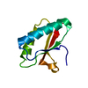
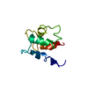
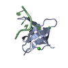
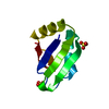
- PDB-4och: Apo structure of Smr domain of MutS2 from Deinococcus radiodurans -
+ Open data

- Basic information
| Entry | Database: PDB / ID: 4och | ||||||
|---|---|---|---|---|---|---|---|
| Title | Apo structure of Smr domain of MutS2 from Deinococcus radiodurans | ||||||
 Components Components | Endonuclease MutS2 | ||||||
 Keywords Keywords |  HYDROLASE / nuclease / Mn2+ ion HYDROLASE / nuclease / Mn2+ ion | ||||||
| Function / homology |  Function and homology information Function and homology informationmismatched DNA binding / negative regulation of DNA recombination / ATP-dependent DNA damage sensor activity / mismatch repair / double-stranded DNA binding / endonuclease activity / Hydrolases; Acting on ester bonds / ATP hydrolysis activity / ATP bindingSimilarity search - Function | ||||||
| Biological species |  Deinococcus radiodurans (radioresistant) Deinococcus radiodurans (radioresistant) | ||||||
| Method | X-RAY DIFFRACTION / SYNCHROTRON / MOLECULAR REPLACEMENT / Resolution: 1.4001 Å | ||||||
 Authors Authors | Zhang, H. / Zhao, Y. / Xu, Q. / Hua, Y.J. | ||||||
 Citation Citation | Journal: Dna Repair / Year: 2014 Title: Structural and functional studies of MutS2 from Deinococcus radiodurans. Authors: Zhang, H. / Xu, Q. / Lu, M. / Xu, X. / Wang, Y. / Wang, L. / Zhao, Y. / Hua, Y. | ||||||
| History |
|
- Structure visualization
| Structure viewer | Molecule: MolmilJmol/JSmol |
|---|
- Downloads & links
-Download
| PDBx/mmCIF format | 4och.cif.gz | 64 KB | Display | PDBx/mmCIF format |
|---|---|---|---|---|
| PDB format | pdb4och.ent.gz | 47.7 KB | Display | PDB format |
| PDBx/mmJSON format | 4och.json.gz | Tree view | PDBx/mmJSON format | |
| Others |  Other downloads Other downloads |
-Validation report
| Arichive directory | https://data.pdbj.org/pub/pdb/validation_reports/oc/4ochftp://data.pdbj.org/pub/pdb/validation_reports/oc/4och | HTTPS FTP |
|---|
-Related structure data


-Links
 PDBj
PDBj- Assembly
| Deposited unit | 
| ||||||||
|---|---|---|---|---|---|---|---|---|---|
| 1 |
| ||||||||
| Unit cell |
|
-Components
| #1: Protein | Mass: 9865.203 Da / Num. of mol.: 1 / Fragment: Smr domain Source method: isolated from a genetically manipulated source Source: (gene. exp.) Deinococcus radiodurans (radioresistant)Strain: ATCC 13939 / DSM 20539 / JCM 16871 / LMG 4051 / NBRC 15346 / NCIMB 9279 / R1 / VKM B-1422 Gene: mutS2, DR_1976 / Production host: Escherichia coli (E. coli) / Strain (production host): BL(21)DE3References: UniProt: Q9RSZ3, Hydrolases; Acting on ester bonds |
|---|---|
| #2: Chemical | ChemComp-GOL / Glycerol  Mass: 92.094 Da / Num. of mol.: 1 / Source method: obtained synthetically / Formula: C3H8O3 Mass: 92.094 Da / Num. of mol.: 1 / Source method: obtained synthetically / Formula: C3H8O3 |
| #3: Water | ChemComp-HOH / Water Mass: 18.015 Da / Num. of mol.: 109 / Source method: isolated from a natural source / Formula: H2O Mass: 18.015 Da / Num. of mol.: 109 / Source method: isolated from a natural source / Formula: H2O |
-Experimental details
-Experiment
| Experiment | Method: X-RAY DIFFRACTION / Number of used crystals: 1 |
|---|
- Sample preparation
| Crystal | Density Matthews: 1.83 Å3/Da / Density % sol: 32.7 % |
|---|---|
| Crystal grow | Temperature: 297 K Method: crystal formed spontaneously in protein solution stored at 4 degree pH: 8.8 Details: 30mM NaCl and 20mM Tris-HCl, pH 8.8, crystal formed spontaneously in protein solution stored at 4 degree, temperature 297K |
-Data collection
| Diffraction | Mean temperature: 100 K | ||||||||||||||||||||||||||||||||||||||||||
|---|---|---|---|---|---|---|---|---|---|---|---|---|---|---|---|---|---|---|---|---|---|---|---|---|---|---|---|---|---|---|---|---|---|---|---|---|---|---|---|---|---|---|---|
| Diffraction source | Source: SYNCHROTRON / Site: SSRF  / Beamline: BL17U / Wavelength: 0.9793 Å / Beamline: BL17U / Wavelength: 0.9793 Å | ||||||||||||||||||||||||||||||||||||||||||
| Detector | Type: ADSC QUANTUM 315r / Detector: CCD / Date: Jun 2, 2011 | ||||||||||||||||||||||||||||||||||||||||||
| Radiation | Monochromator: silicon 111 / Protocol: SINGLE WAVELENGTH / Monochromatic (M) / Laue (L): M / Scattering type: x-ray | ||||||||||||||||||||||||||||||||||||||||||
| Radiation wavelength | Wavelength: 0.9793 Å / Relative weight: 1 | ||||||||||||||||||||||||||||||||||||||||||
| Reflection | Resolution: 1.4→30 Å / Num. all: 14061 / Num. obs: 13788 / % possible obs: 98.1 % / Observed criterion σ(I): -3 / Redundancy: 3.6 % / Rmerge(I) obs: 0.07 / Net I/σ(I): 14.02 | ||||||||||||||||||||||||||||||||||||||||||
| Reflection shell |
|
- Processing
| Software |
| ||||||||||||||||||||||||||||||||||||||||||
|---|---|---|---|---|---|---|---|---|---|---|---|---|---|---|---|---|---|---|---|---|---|---|---|---|---|---|---|---|---|---|---|---|---|---|---|---|---|---|---|---|---|---|---|
| Refinement | Method to determine structure: MOLECULAR REPLACEMENT / Resolution: 1.4001→25.598 Å / SU ML: 0.16 / σ(F): 1.34 / Phase error: 24.77 / Stereochemistry target values: ML
| ||||||||||||||||||||||||||||||||||||||||||
| Solvent computation | Shrinkage radii: 0.9 Å / VDW probe radii: 1.11 Å / Solvent model: FLAT BULK SOLVENT MODEL | ||||||||||||||||||||||||||||||||||||||||||
| Refinement step | Cycle: LAST / Resolution: 1.4001→25.598 Å
| ||||||||||||||||||||||||||||||||||||||||||
| Refine LS restraints |
| ||||||||||||||||||||||||||||||||||||||||||
| LS refinement shell |
|
