[English] 日本語
Yorodumi
- PDB-4o1p: Crystal Structure of RNase L in complex with 2-5A and AMP-PNP -

+
Open data


ID or keywords:

Loading...

-
Basic information

Entry
Database: PDB / ID: 4o1p
TitleCrystal Structure of RNase L in complex with 2-5A and AMP-PNP
ComponentsRibonuclease L
KeywordsTRANSFERASE / HYDROLASE / ankyrin repeat-kinase-RNAse / RNA cleavage / 2-5A
Function / homology
Function and homology information


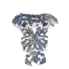
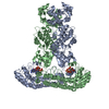
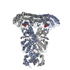
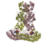
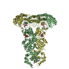
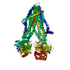
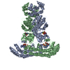
regulation of RNA metabolic process / negative regulation of viral genome replication / RNA nuclease activity / mRNA processing / defense response to virus / protein kinase activity / RNA binding / ATP binding / metal ion binding
Similarity search - Function
RNase L, RNase domain / KEN domain / KEN domain / KEN domain superfamily / Ribonuclease 2-5A / KEN domain profile. / domain in protein kinases, N-glycanases and other nuclear proteins / de novo design (two linked rop proteins) / Ankyrin repeat-containing domain / Ankyrin repeat ...RNase L, RNase domain / KEN domain / KEN domain / KEN domain superfamily / Ribonuclease 2-5A / KEN domain profile. / domain in protein kinases, N-glycanases and other nuclear proteins / de novo design (two linked rop proteins) / Ankyrin repeat-containing domain / Ankyrin repeat / Ankyrin repeats (3 copies) / Ankyrin repeat profile. / Ankyrin repeat region circular profile. / ankyrin repeats / Ankyrin repeat / Ankyrin repeat-containing domain superfamily / Serine Threonine Protein Phosphatase 5, Tetratricopeptide repeat / Alpha Horseshoe / Transferase(Phosphotransferase) domain 1 / Transferase(Phosphotransferase); domain 1 / Protein kinase domain / Serine/Threonine protein kinases, catalytic domain / Protein kinase domain profile. / Protein kinase domain / Protein kinase-like domain superfamily / Up-down Bundle / Orthogonal Bundle / Mainly Alpha
Similarity search - Domain/homology
Chem-25L / PHOSPHOAMINOPHOSPHONIC ACID-ADENYLATE ESTER / Ribonuclease L
Similarity search - Component
Biological speciesSus scrofa (pig)
MethodX-RAY DIFFRACTION / SYNCHROTRON / molecular replacement-SAD / Resolution: 2.5 Å
AuthorsHuang, H. / Zeqiraj, E. / Ceccarelli, D.F. / Sicheri, F.
CitationJournal: Mol.Cell / Year: 2014
Title: Dimeric structure of pseudokinase RNase L bound to 2-5A reveals a basis for interferon-induced antiviral activity.
Authors: Huang, H. / Zeqiraj, E. / Dong, B. / Jha, B.K. / Duffy, N.M. / Orlicky, S. / Thevakumaran, N. / Talukdar, M. / Pillon, M.C. / Ceccarelli, D.F. / Wan, L.C. / Juang, Y.C. / Mao, D.Y. / ...Authors: Huang, H. / Zeqiraj, E. / Dong, B. / Jha, B.K. / Duffy, N.M. / Orlicky, S. / Thevakumaran, N. / Talukdar, M. / Pillon, M.C. / Ceccarelli, D.F. / Wan, L.C. / Juang, Y.C. / Mao, D.Y. / Gaughan, C. / Brinton, M.A. / Perelygin, A.A. / Kourinov, I. / Guarne, A. / Silverman, R.H. / Sicheri, F.
History
DepositionDec 16, 2013Deposition site: RCSB / Processing site: RCSB
Revision 1.0Feb 5, 2014Provider: repository / Type: Initial release
Revision 1.1Dec 24, 2014Group: Database references
Revision 1.2Sep 20, 2023Group: Data collection / Database references ...Data collection / Database references / Derived calculations / Refinement description
Category: chem_comp_atom / chem_comp_bond ...chem_comp_atom / chem_comp_bond / database_2 / pdbx_initial_refinement_model / pdbx_struct_conn_angle / struct_conn / struct_ncs_dom_lim / struct_ref_seq_dif / struct_site
Item: _database_2.pdbx_DOI / _database_2.pdbx_database_accession ..._database_2.pdbx_DOI / _database_2.pdbx_database_accession / _pdbx_struct_conn_angle.ptnr1_auth_asym_id / _pdbx_struct_conn_angle.ptnr1_auth_comp_id / _pdbx_struct_conn_angle.ptnr1_auth_seq_id / _pdbx_struct_conn_angle.ptnr1_label_asym_id / _pdbx_struct_conn_angle.ptnr1_label_atom_id / _pdbx_struct_conn_angle.ptnr1_label_comp_id / _pdbx_struct_conn_angle.ptnr1_label_seq_id / _pdbx_struct_conn_angle.ptnr2_auth_asym_id / _pdbx_struct_conn_angle.ptnr2_auth_seq_id / _pdbx_struct_conn_angle.ptnr2_label_asym_id / _pdbx_struct_conn_angle.ptnr3_auth_asym_id / _pdbx_struct_conn_angle.ptnr3_auth_comp_id / _pdbx_struct_conn_angle.ptnr3_auth_seq_id / _pdbx_struct_conn_angle.ptnr3_label_asym_id / _pdbx_struct_conn_angle.ptnr3_label_atom_id / _pdbx_struct_conn_angle.ptnr3_label_comp_id / _pdbx_struct_conn_angle.ptnr3_label_seq_id / _pdbx_struct_conn_angle.value / _struct_conn.pdbx_dist_value / _struct_conn.ptnr1_auth_asym_id / _struct_conn.ptnr1_auth_comp_id / _struct_conn.ptnr1_auth_seq_id / _struct_conn.ptnr1_label_asym_id / _struct_conn.ptnr1_label_atom_id / _struct_conn.ptnr1_label_comp_id / _struct_conn.ptnr1_label_seq_id / _struct_conn.ptnr2_auth_asym_id / _struct_conn.ptnr2_auth_comp_id / _struct_conn.ptnr2_auth_seq_id / _struct_conn.ptnr2_label_asym_id / _struct_conn.ptnr2_label_atom_id / _struct_conn.ptnr2_label_comp_id / _struct_ncs_dom_lim.beg_auth_comp_id / _struct_ncs_dom_lim.beg_label_asym_id / _struct_ncs_dom_lim.beg_label_comp_id / _struct_ncs_dom_lim.beg_label_seq_id / _struct_ncs_dom_lim.end_auth_comp_id / _struct_ncs_dom_lim.end_label_asym_id / _struct_ncs_dom_lim.end_label_comp_id / _struct_ncs_dom_lim.end_label_seq_id / _struct_ref_seq_dif.details / _struct_site.pdbx_auth_asym_id / _struct_site.pdbx_auth_comp_id / _struct_site.pdbx_auth_seq_id

-
Structure visualization

Structure viewerMolecule:
MolmilJmol/JSmol

Downloads & links

-
Assembly

Deposited unit
A: Ribonuclease L
B: Ribonuclease L
C: Ribonuclease L
D: Ribonuclease L
hetero molecules


Theoretical massNumber of molelcules
Total (without water)332,19320
Polymers325,3114
Non-polymers6,88216
Water18,5551030
1
A: Ribonuclease L
B: Ribonuclease L
hetero molecules


Theoretical massNumber of molelcules
Total (without water)166,09610
Polymers162,6562
Non-polymers3,4418
Water362
TypeNameSymmetry operationNumber
identity operation1_555x,y,z1
Buried area7120 Å2
ΔGint13 kcal/mol
Surface area58630 Å2
MethodPISA
2
C: Ribonuclease L
D: Ribonuclease L
hetero molecules


Theoretical massNumber of molelcules
Total (without water)166,09610
Polymers162,6562
Non-polymers3,4418
Water362
TypeNameSymmetry operationNumber
identity operation1_555x,y,z1
Buried area6960 Å2
ΔGint13 kcal/mol
Surface area58570 Å2
MethodPISA
Unit cell
Length a, b, c (Å)59.197, 267.392, 110.210
Angle α, β, γ (deg.)90.00, 90.04, 90.00
Int Tables number4
Space group name H-MP1211
Noncrystallographic symmetry (NCS)NCS domain:
IDEns-IDDetails
11A
21B
12A
22C
13A
23D
14B
24C
15B
25D
16C
26D

NCS domain segments:

Component-ID: 0 / Beg auth comp-ID: SER / Beg label comp-ID: SER / End auth comp-ID: ASN / End label comp-ID: ASN / Refine code: 0 / Auth seq-ID: 22 - 729 / Label seq-ID: 7 - 714

Dom-IDEns-IDAuth asym-IDLabel asym-ID
11AA
21BB
12AA
22CC
13AA
23DD
14BB
24CC
15BB
25DD
16CC
26DD

NCS ensembles :
ID
1
2
3
4
5
6

-
Components

#1: Protein
Ribonuclease L /


Mass: 81327.836 Da / Num. of mol.: 4 / Fragment: UNP residues 21-732
Source method: isolated from a genetically manipulated source
Source: (gene. exp.) Sus scrofa (pig) / Gene: RNASEL / Production host: Escherichia coli (E. coli) / References: UniProt: A5H025
#2: Chemical
ChemComp-ANP / PHOSPHOAMINOPHOSPHONIC ACID-ADENYLATE ESTER


Mass: 506.196 Da / Num. of mol.: 4 / Source method: obtained synthetically / Formula: C10H17N6O12P3 / Comment: AMP-PNP, energy-carrying molecule analogue*YM
#3: Chemical
ChemComp-MG / MAGNESIUM ION


Mass: 24.305 Da / Num. of mol.: 8 / Source method: obtained synthetically / Formula: Mg
#4: Chemical
ChemComp-25L / [[(2R,3R,4R,5R)-5-(6-aminopurin-9-yl)-4-[[(2R,3R,4R,5R)-5-(6-aminopurin-9-yl)-4-[[(2R,3S,4R,5R)-5-(6-aminopurin-9-yl)-3,4-dihydroxy-oxolan-2-yl]methoxy-hydroxy-phosphoryl]oxy-3-hydroxy-oxolan-2-yl]methoxy-hydroxy-phosphoryl]oxy-3-hydroxy-oxolan-2-yl]methoxy-hydroxy-phosphoryl] phosphono hydrogen phosphate / 2'-5'-oligoadenylate trimer


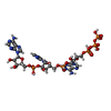
Mass: 1165.593 Da / Num. of mol.: 4 / Source method: obtained synthetically / Formula: C30H40N15O25P5
#5: Water ChemComp-HOH / water / Water


Mass: 18.015 Da / Num. of mol.: 1030 / Source method: isolated from a natural source / Formula: H2O

-
Experimental details

-
Experiment

ExperimentMethod: X-RAY DIFFRACTION / Number of used crystals: 1

-
Sample preparation

CrystalDensity Matthews: 2.68 Å3/Da / Density % sol: 54.13 %
Crystal growTemperature: 293 K / Method: vapor diffusion, hanging drop / pH: 7
Details: 5 mM MgCl2, 5 mM DTT, 18% PEG2000, 100 mM NaCl, and 100 mM SPG buffer, pH 7. , VAPOR DIFFUSION, HANGING DROP, temperature 293K

-
Data collection

DiffractionMean temperature: 100 K
Diffraction sourceSource: SYNCHROTRON / Site: APS / Beamline: 24-ID-C / Wavelength: 0.9792 Å
DetectorType: DECTRIS PILATUS 6M / Detector: PIXEL / Date: Feb 1, 2013
RadiationMonochromator: SI(111) / Protocol: SINGLE WAVELENGTH / Monochromatic (M) / Laue (L): M / Scattering type: x-ray
Radiation wavelengthWavelength: 0.9792 Å / Relative weight: 1
Reflection twin
Crystal-IDIDOperatorDomain-IDFraction
11H, K, L10.773
11h,-k,-l20.227
ReflectionResolution: 2.5→59.2 Å / Num. obs: 106347 / % possible obs: 95.7 % / Observed criterion σ(F): 1.6 / Observed criterion σ(I): 1.6 / Redundancy: 3 % / Rmerge(I) obs: 0.104 / Net I/σ(I): 6.8
Reflection shellResolution: 2.504→2.569 Å / Redundancy: 3.2 % / Rmerge(I) obs: 0.526 / Mean I/σ(I) obs: 1.6 / Num. unique all: 7277 / % possible all: 88.4

-
Processing

Software
NameVersionClassification
ADSCQuantumdata collection
PHASERphasing
REFMAC5.7.0032refinement
XDSdata reduction
XDSdata scaling
RefinementMethod to determine structure: molecular replacement-SAD
Starting model: PDB ENTRY 4G8K
Resolution: 2.5→59.2 Å / Cor.coef. Fo:Fc: 0.938 / Cor.coef. Fo:Fc free: 0.918 / SU B: 7.961 / SU ML: 0.185 / Cross valid method: THROUGHOUT / σ(F): 1.6 / ESU R: 0.133 / ESU R Free: 0.059 / Stereochemistry target values: MAXIMUM LIKELIHOOD / Details: HYDROGENS HAVE BEEN ADDED IN THE RIDING POSITIONS
RfactorNum. reflection% reflectionSelection details
Rfree0.23107 5725 5.1 %RANDOM
Rwork0.19568 ---
obs0.1975 106347 95.67 %-
all-111125 --
Solvent computationIon probe radii: 0.8 Å / Shrinkage radii: 0.8 Å / VDW probe radii: 1.2 Å / Solvent model: MASK
Displacement parametersBiso mean: 51.977 Å2
Baniso -1Baniso -2Baniso -3
1-2.95 Å20 Å21.86 Å2
2---18.36 Å20 Å2
3---15.41 Å2
Refinement stepCycle: LAST / Resolution: 2.5→59.2 Å
ProteinNucleic acidLigandSolventTotal
Num. atoms21367 0 432 1030 22829
Refine LS restraints
Refine-IDTypeDev idealDev ideal targetNumber
X-RAY DIFFRACTIONr_bond_refined_d0.0070.01922220
X-RAY DIFFRACTIONr_bond_other_d0.0030.0220975
X-RAY DIFFRACTIONr_angle_refined_deg1.2311.98230092
X-RAY DIFFRACTIONr_angle_other_deg0.8323.00148204
X-RAY DIFFRACTIONr_dihedral_angle_1_deg5.39452666
X-RAY DIFFRACTIONr_dihedral_angle_2_deg36.04424.7121112
X-RAY DIFFRACTIONr_dihedral_angle_3_deg16.195153934
X-RAY DIFFRACTIONr_dihedral_angle_4_deg18.38615152
X-RAY DIFFRACTIONr_chiral_restr0.0730.23322
X-RAY DIFFRACTIONr_gen_planes_refined0.0040.0225018
X-RAY DIFFRACTIONr_gen_planes_other0.0030.025114
X-RAY DIFFRACTIONr_mcbond_it2.6955.10210724
X-RAY DIFFRACTIONr_mcbond_other2.6925.10110723
X-RAY DIFFRACTIONr_mcangle_it4.3577.63813370
X-RAY DIFFRACTIONr_mcangle_other4.3577.63813371
X-RAY DIFFRACTIONr_scbond_it2.7895.33611496
X-RAY DIFFRACTIONr_scbond_other2.7885.33611490
X-RAY DIFFRACTIONr_scangle_other4.6167.88516713
X-RAY DIFFRACTIONr_long_range_B_refined9.06947.46693506
X-RAY DIFFRACTIONr_long_range_B_other9.06547.47993190
Refine LS restraints NCS

Refine-ID: X-RAY DIFFRACTION / Type: interatomic distance / Weight position: 0.05

Ens-IDDom-IDAuth asym-IDNumberRms dev position (Å)
11A427870.04
12B427870.04
21A430520.03
22C430520.03
31A425260.04
32D425260.04
41B429760.02
42C429760.02
51B428440.03
52D428440.03
61C426870.03
62D426870.03
LS refinement shellResolution: 2.504→2.569 Å / Total num. of bins used: 20
RfactorNum. reflection% reflection
Rfree0.304 403 -
Rwork0.273 7277 -
obs--88.4 %

+
About Yorodumi

-
News

-
Feb 9, 2022. New format data for meta-information of EMDB entries

New format data for meta-information of EMDB entries

  • Version 3 of the EMDB header file is now the official format.
  • The previous official version 1.9 will be removed from the archive.

Related info.:EMDB header

External links:wwPDB to switch to version 3 of the EMDB data model

-
Aug 12, 2020. Covid-19 info

Covid-19 info

URL: https://pdbj.org/emnavi/covid19.php

New page: Covid-19 featured information page in EM Navigator.

Related info.:Covid-19 info / Mar 5, 2020. Novel coronavirus structure data

+
Mar 5, 2020. Novel coronavirus structure data

Novel coronavirus structure data

Related info.:Yorodumi Speices / Aug 12, 2020. Covid-19 info

External links:COVID-19 featured content - PDBj / Molecule of the Month (242):Coronavirus Proteases

+
Jan 31, 2019. EMDB accession codes are about to change! (news from PDBe EMDB page)

EMDB accession codes are about to change! (news from PDBe EMDB page)

  • The allocation of 4 digits for EMDB accession codes will soon come to an end. Whilst these codes will remain in use, new EMDB accession codes will include an additional digit and will expand incrementally as the available range of codes is exhausted. The current 4-digit format prefixed with “EMD-” (i.e. EMD-XXXX) will advance to a 5-digit format (i.e. EMD-XXXXX), and so on. It is currently estimated that the 4-digit codes will be depleted around Spring 2019, at which point the 5-digit format will come into force.
  • The EM Navigator/Yorodumi systems omit the EMD- prefix.

Related info.:Q: What is EMD? / ID/Accession-code notation in Yorodumi/EM Navigator

External links:EMDB Accession Codes are Changing Soon! / Contact to PDBj

+
Jul 12, 2017. Major update of PDB

Major update of PDB

  • wwPDB released updated PDB data conforming to the new PDBx/mmCIF dictionary.
  • This is a major update changing the version number from 4 to 5, and with Remediation, in which all the entries are updated.
  • In this update, many items about electron microscopy experimental information are reorganized (e.g. em_software).
  • Now, EM Navigator and Yorodumi are based on the updated data.

External links:wwPDB Remediation / Enriched Model Files Conforming to OneDep Data Standards Now Available in the PDB FTP Archive

-
Yorodumi

Thousand views of thousand structures

  • Yorodumi is a browser for structure data from EMDB, PDB, SASBDB, etc.
  • This page is also the successor to EM Navigator detail page, and also detail information page/front-end page for Omokage search.
  • The word "yorodu" (or yorozu) is an old Japanese word meaning "ten thousand". "mi" (miru) is to see.

Related info.:EMDB / PDB / SASBDB / Comparison of 3 databanks / Yorodumi Search / Aug 31, 2016. New EM Navigator & Yorodumi / Yorodumi Papers / Jmol/JSmol / Function and homology information / Changes in new EM Navigator and Yorodumi

Read more