 Movie
Movie Controller

[English] 日本語
 Yorodumi
- PDB-4nvp: Structure of the cyclic nucleotide-binding domain of HCN4 channel... -
+ Open data

- Basic information
| Entry | Database: PDB / ID: 4nvp | ||||||
|---|---|---|---|---|---|---|---|
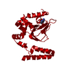
| Title | Structure of the cyclic nucleotide-binding domain of HCN4 channel complexed with 7-CH-cAMP | ||||||
 Components Components | Potassium/sodium hyperpolarization-activated cyclic nucleotide-gated channel 4 | ||||||
 Keywords Keywords |  TRANSPORT PROTEIN / cyclic nucleotide binding domain / Potassium/sodium hyperpolarization-activated cyclic nucleotide-gated channel / cytoplasmic domain TRANSPORT PROTEIN / cyclic nucleotide binding domain / Potassium/sodium hyperpolarization-activated cyclic nucleotide-gated channel / cytoplasmic domain | ||||||
| Function / homology |  Function and homology information Function and homology informationvoltage-gated potassium channel activity involved in SA node cell action potential depolarization / sinoatrial node development / HCN channels / HCN channel complex / regulation of cardiac muscle cell action potential involved in regulation of contraction / SA node cell action potential / membrane depolarization during SA node cell action potential / intracellularly cAMP-activated cation channel activity / cellular response to cGMP / membrane depolarization during cardiac muscle cell action potential ...voltage-gated potassium channel activity involved in SA node cell action potential depolarization / sinoatrial node development / HCN channels / HCN channel complex / regulation of cardiac muscle cell action potential involved in regulation of contraction / SA node cell action potential / membrane depolarization during SA node cell action potential / intracellularly cAMP-activated cation channel activity / cellular response to cGMP / membrane depolarization during cardiac muscle cell action potential / sodium ion import across plasma membrane / voltage-gated sodium channel activity / blood circulation / regulation of membrane depolarization / potassium ion import across plasma membrane / regulation of heart rate by cardiac conduction / voltage-gated potassium channel activity / monoatomic cation transport / sodium ion transmembrane transport / regulation of cardiac muscle contraction / cAMP binding / cellular response to cAMP / potassium ion transmembrane transport / regulation of heart rate / regulation of membrane potential / muscle contraction / axon / dendrite / perinuclear region of cytoplasm / identical protein binding / plasma membraneSimilarity search - Function | ||||||
| Biological species |  Homo sapiens (human) Homo sapiens (human) | ||||||
| Method | X-RAY DIFFRACTION / SYNCHROTRON / MOLECULAR REPLACEMENT / Resolution: 2.5 Å | ||||||
 Authors Authors | Alfieri, A. / Moroni, A. | ||||||
 Citation Citation | Journal: Acs Chem.Biol. / Year: 2014 Title: Cyclic Nucleotide Mapping of Hyperpolarization-Activated Cyclic Nucleotide-Gated (HCN) Channels. Authors: Moller, S. / Alfieri, A. / Bertinetti, D. / Aquila, M. / Schwede, F. / Lolicato, M. / Rehmann, H. / Moroni, A. / Herberg, F.W. | ||||||
| History |
|
- Structure visualization
| Structure viewer | Molecule: MolmilJmol/JSmol |
|---|
- Downloads & links
-Download
| PDBx/mmCIF format | 4nvp.cif.gz | 56.5 KB | Display | PDBx/mmCIF format |
|---|---|---|---|---|
| PDB format | pdb4nvp.ent.gz | 40.7 KB | Display | PDB format |
| PDBx/mmJSON format | 4nvp.json.gz | Tree view | PDBx/mmJSON format | |
| Others |  Other downloads Other downloads |
-Validation report
| Arichive directory | https://data.pdbj.org/pub/pdb/validation_reports/nv/4nvpftp://data.pdbj.org/pub/pdb/validation_reports/nv/4nvp | HTTPS FTP |
|---|
-Related structure data
| Related structure data | |
|---|---|
| Similar structure data |


-Links
 PDBj
PDBj


- Assembly
| Deposited unit | 
| ||||||||
|---|---|---|---|---|---|---|---|---|---|
| 1 | 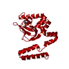
| ||||||||
| Unit cell |
| ||||||||
| Components on special symmetry positions |
|
-Components
| #1: Protein | Mass: 24498.004 Da / Num. of mol.: 1 Source method: isolated from a genetically manipulated source Source: (gene. exp.) Homo sapiens (human) / Gene: HCN4 / Production host:  Escherichia coli (E. coli) / Strain (production host): BL21(DE3) / References: UniProt: Q9Y3Q4 Escherichia coli (E. coli) / Strain (production host): BL21(DE3) / References: UniProt: Q9Y3Q4 |
|---|---|
| #2: Chemical | ChemComp-7CH / (  Mass: 328.218 Da / Num. of mol.: 1 / Source method: obtained synthetically / Formula: C11H13N4O6P Mass: 328.218 Da / Num. of mol.: 1 / Source method: obtained synthetically / Formula: C11H13N4O6P |
| #3: Water | ChemComp-HOH / Water Mass: 18.015 Da / Num. of mol.: 37 / Source method: isolated from a natural source / Formula: H2O Mass: 18.015 Da / Num. of mol.: 37 / Source method: isolated from a natural source / Formula: H2O |
-Experimental details
-Experiment
| Experiment | Method: X-RAY DIFFRACTION / Number of used crystals: 1 |
|---|
- Sample preparation
| Crystal | Density Matthews: 2.38 Å3/Da / Density % sol: 48.39 % |
|---|---|
| Crystal grow | Temperature: 277 K / Method: vapor diffusion, sitting drop / pH: 8 Details: HCN4 521-723 (10 mg mL-1) and 7-CH-cAMP 0.5mM were mixed in a 2:1 ratio with 100 mM TrisCl pH 8.0, 20 % PEG4000 (w/v), VAPOR DIFFUSION, SITTING DROP, temperature 277K |
-Data collection
| Diffraction | Mean temperature: 100 K |
|---|---|
| Diffraction source | Source: SYNCHROTRON / Site: ESRF  / Beamline: BM14 / Wavelength: 0.97372 Å / Beamline: BM14 / Wavelength: 0.97372 Å |
| Detector | Type: MARMOSAIC 225 mm CCD / Detector: CCD / Date: Oct 24, 2013 / Details: bending magnet; bent collimating mirror and toroid |
| Radiation | Monochromator: Si(111) monochromator / Protocol: SINGLE WAVELENGTH / Monochromatic (M) / Laue (L): M / Scattering type: x-ray |
| Radiation wavelength | Wavelength: 0.97372 Å / Relative weight: 1 |
| Reflection | Resolution: 2.5→50.17 Å / Num. all: 8568 / Num. obs: 8568 / % possible obs: 100 % / Observed criterion σ(I): 5 / Redundancy: 8.1 % / Biso Wilson estimate: 33.2 Å2 / Rmerge(I) obs: 0.08 / Net I/σ(I): 16.4 |
| Reflection shell | Resolution: 2.5→2.64 Å / Redundancy: 7.7 % / Rmerge(I) obs: 0.45 / Mean I/σ(I) obs: 4.3 / Num. unique all: 1215 / % possible all: 99.9 |
- Processing
| Software |
| |||||||||||||||||||||||||||||||||||||||||||||||||||||||||||||||||||||||||||||||||||||||||||||||||||||||||
|---|---|---|---|---|---|---|---|---|---|---|---|---|---|---|---|---|---|---|---|---|---|---|---|---|---|---|---|---|---|---|---|---|---|---|---|---|---|---|---|---|---|---|---|---|---|---|---|---|---|---|---|---|---|---|---|---|---|---|---|---|---|---|---|---|---|---|---|---|---|---|---|---|---|---|---|---|---|---|---|---|---|---|---|---|---|---|---|---|---|---|---|---|---|---|---|---|---|---|---|---|---|---|---|---|---|---|
| Refinement | Method to determine structure: MOLECULAR REPLACEMENT Starting model: PDB ENTRY 3U11_A Resolution: 2.5→44.47 Å / Cor.coef. Fo:Fc: 0.942 / Cor.coef. Fo:Fc free: 0.919 / SU B: 9.961 / SU ML: 0.221 / Cross valid method: THROUGHOUT / ESU R: 0.604 / ESU R Free: 0.317 / Stereochemistry target values: MAXIMUM LIKELIHOOD / Details: HYDROGENS HAVE BEEN ADDED IN THE RIDING POSITIONS
| |||||||||||||||||||||||||||||||||||||||||||||||||||||||||||||||||||||||||||||||||||||||||||||||||||||||||
| Solvent computation | Ion probe radii: 0.8 Å / Shrinkage radii: 0.8 Å / VDW probe radii: 1.2 Å / Solvent model: MASK | |||||||||||||||||||||||||||||||||||||||||||||||||||||||||||||||||||||||||||||||||||||||||||||||||||||||||
| Displacement parameters | Biso mean: 42.441 Å2
| |||||||||||||||||||||||||||||||||||||||||||||||||||||||||||||||||||||||||||||||||||||||||||||||||||||||||
| Refinement step | Cycle: LAST / Resolution: 2.5→44.47 Å
| |||||||||||||||||||||||||||||||||||||||||||||||||||||||||||||||||||||||||||||||||||||||||||||||||||||||||
| Refine LS restraints |
| |||||||||||||||||||||||||||||||||||||||||||||||||||||||||||||||||||||||||||||||||||||||||||||||||||||||||
| LS refinement shell | Resolution: 2.5→2.565 Å / Total num. of bins used: 20
|
