 Movie
Movie Controller

[English] 日本語
 Yorodumi
- PDB-4nup: Crystal structure of mouse N-cadherin EC1-2 with AA insertion bet... -
+ Open data

- Basic information
| Entry | Database: PDB / ID: 4nup | ||||||
|---|---|---|---|---|---|---|---|
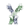
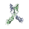
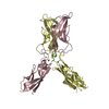
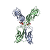
| Title | Crystal structure of mouse N-cadherin EC1-2 with AA insertion between residues 2 and 3 | ||||||
 Components Components | N-CADHERIN EC1-2 | ||||||
 Keywords Keywords |  CELL ADHESION / cell adhesion molecule CELL ADHESION / cell adhesion molecule | ||||||
| Function / homology |  Function and homology information Function and homology informationmesenchymal cell migration / regulation of oligodendrocyte progenitor proliferation / regulation of postsynaptic density protein 95 clustering / radial glial cell differentiation / neuroligin clustering involved in postsynaptic membrane assembly / positive regulation of synaptic vesicle clustering / Regulation of Insulin-like Growth Factor (IGF) transport and uptake by Insulin-like Growth Factor Binding Proteins (IGFBPs) / Post-translational protein phosphorylation / Adherens junctions interactions / desmosome ...mesenchymal cell migration / regulation of oligodendrocyte progenitor proliferation / regulation of postsynaptic density protein 95 clustering / radial glial cell differentiation / neuroligin clustering involved in postsynaptic membrane assembly / positive regulation of synaptic vesicle clustering / Regulation of Insulin-like Growth Factor (IGF) transport and uptake by Insulin-like Growth Factor Binding Proteins (IGFBPs) / Post-translational protein phosphorylation / Adherens junctions interactions / desmosome / synaptic vesicle clustering / gamma-catenin binding / neural crest cell development / glial cell differentiation / telencephalon development / neuroepithelial cell differentiation / type B pancreatic cell development / cell-cell adhesion mediated by cadherin / neuronal stem cell population maintenance / alpha-catenin binding / fascia adherens / apicolateral plasma membrane / calcium-dependent cell-cell adhesion via plasma membrane cell adhesion molecules / Myogenesis / regulation of Rho protein signal transduction / postsynaptic specialization membrane / brain morphogenesis / catenin complex / cell-cell junction assembly / adherens junction organization / blood vessel morphogenesis / regulation of myelination / heterophilic cell-cell adhesion via plasma membrane cell adhesion molecules / regulation of axonogenesis / cortical actin cytoskeleton / nitric-oxide synthase binding / homophilic cell adhesion via plasma membrane adhesion molecules / plasma membrane raft / intercalated disc / homeostasis of number of cells / regulation of signal transduction / presynaptic active zone membrane / regulation of synaptic transmission, glutamatergic / T-tubule / synapse assembly / striated muscle cell differentiation / protein tyrosine kinase binding / protein localization to plasma membrane / adherens junction / sarcolemma / brain development / modulation of chemical synaptic transmission / cell morphogenesis / negative regulation of canonical Wnt signaling pathway / cerebral cortex development / cell-cell adhesion / beta-catenin binding / regulation of protein localization / cell migration / cell-cell junction / apical part of cell / cell junction / lamellipodium / basolateral plasma membrane / protein phosphatase binding / positive regulation of MAPK cascade / postsynaptic density / membrane => GO:0016020 / cell adhesion / neuron projection / cadherin binding / apical plasma membrane / synapse / glutamatergic synapse / calcium ion binding / protein-containing complex binding / protein kinase binding / enzyme binding / cell surface / protein-containing complex / RNA binding / membrane / identical protein binding / plasma membrane / cytoplasmSimilarity search - Function | ||||||
| Biological species |  Mus musculus (house mouse) Mus musculus (house mouse) | ||||||
| Method | X-RAY DIFFRACTION / SYNCHROTRON / MOLECULAR REPLACEMENT / Resolution: 2.7 Å | ||||||
 Authors Authors | Jin, X. | ||||||
 Citation Citation | Journal: Proc.Natl.Acad.Sci.USA / Year: 2014 Title: Structural and energetic determinants of adhesive binding specificity in type I cadherins. Authors: Vendome, J. / Felsovalyi, K. / Song, H. / Yang, Z. / Jin, X. / Brasch, J. / Harrison, O.J. / Ahlsen, G. / Bahna, F. / Kaczynska, A. / Katsamba, P.S. / Edmond, D. / Hubbell, W.L. / Shapiro, L. / Honig, B. | ||||||
| History |
|
- Structure visualization
| Structure viewer | Molecule: MolmilJmol/JSmol |
|---|
- Downloads & links
-Download
| PDBx/mmCIF format | 4nup.cif.gz | 152.2 KB | Display | PDBx/mmCIF format |
|---|---|---|---|---|
| PDB format | pdb4nup.ent.gz | 119.6 KB | Display | PDB format |
| PDBx/mmJSON format | 4nup.json.gz | Tree view | PDBx/mmJSON format | |
| Others |  Other downloads Other downloads |
-Validation report
| Arichive directory | https://data.pdbj.org/pub/pdb/validation_reports/nu/4nupftp://data.pdbj.org/pub/pdb/validation_reports/nu/4nup | HTTPS FTP |
|---|
-Related structure data


-Links
 PDBj
PDBj
- Assembly
| Deposited unit | 
| ||||||||
|---|---|---|---|---|---|---|---|---|---|
| 1 | 
| ||||||||
| 2 | 
| ||||||||
| Unit cell |
|
-Components
| #1: Protein | Mass: 23759.646 Da / Num. of mol.: 3 / Fragment: unp residues 160-374 Source method: isolated from a genetically manipulated source Source: (gene. exp.) Mus musculus (house mouse) / Gene: Cdh2 / Production host:  Escherichia coli (E. coli) / References: UniProt: Q3UIC2, UniProt: P15116*PLUS Escherichia coli (E. coli) / References: UniProt: Q3UIC2, UniProt: P15116*PLUS#2: Chemical | ChemComp-CA /   Mass: 40.078 Da / Num. of mol.: 10 / Source method: obtained synthetically / Formula: Ca Mass: 40.078 Da / Num. of mol.: 10 / Source method: obtained synthetically / Formula: Ca#3: Water | ChemComp-HOH / | Water Mass: 18.015 Da / Num. of mol.: 576 / Source method: isolated from a natural source / Formula: H2O Mass: 18.015 Da / Num. of mol.: 576 / Source method: isolated from a natural source / Formula: H2O |
|---|
-Experimental details
-Experiment
| Experiment | Method: X-RAY DIFFRACTION / Number of used crystals: 1 |
|---|
- Sample preparation
| Crystal | Density Matthews: 4.15 Å3/Da / Density % sol: 70.37 % |
|---|---|
| Crystal grow | Temperature: 293 K / Method: vapor diffusion, hanging drop / pH: 7.5 Details: 0.3M magnesium formate, 0.1M HEPES, pH 7.5, VAPOR DIFFUSION, HANGING DROP, temperature 293K |
-Data collection
| Diffraction | Mean temperature: 100 K |
|---|---|
| Diffraction source | Source: SYNCHROTRON / Site: NSLS  / Beamline: X4C / Wavelength: 0.9793 Å / Beamline: X4C / Wavelength: 0.9793 Å |
| Detector | Type: MAR CCD 165 mm / Detector: CCD / Date: Sep 9, 2010 |
| Radiation | Monochromator: SI 111 CHANNEL / Protocol: SINGLE WAVELENGTH / Monochromatic (M) / Laue (L): M / Scattering type: x-ray |
| Radiation wavelength | Wavelength: 0.9793 Å / Relative weight: 1 |
| Reflection | Resolution: 2.7→20 Å / Num. all: 34109 / Num. obs: 33779 / % possible obs: 99 % / Observed criterion σ(F): 1 / Observed criterion σ(I): 1 |
- Processing
| Software |
| |||||||||||||||||||||||||||||||||||||||||||||||||||||||||||||||||||||||||||||||||||||||||||
|---|---|---|---|---|---|---|---|---|---|---|---|---|---|---|---|---|---|---|---|---|---|---|---|---|---|---|---|---|---|---|---|---|---|---|---|---|---|---|---|---|---|---|---|---|---|---|---|---|---|---|---|---|---|---|---|---|---|---|---|---|---|---|---|---|---|---|---|---|---|---|---|---|---|---|---|---|---|---|---|---|---|---|---|---|---|---|---|---|---|---|---|---|
| Refinement | Method to determine structure: MOLECULAR REPLACEMENT / Resolution: 2.7→19.963 Å / SU ML: 0.3 / σ(F): 1.34 / Phase error: 22.2 / Stereochemistry target values: ML
| |||||||||||||||||||||||||||||||||||||||||||||||||||||||||||||||||||||||||||||||||||||||||||
| Solvent computation | Shrinkage radii: 0.9 Å / VDW probe radii: 1.11 Å / Solvent model: FLAT BULK SOLVENT MODEL | |||||||||||||||||||||||||||||||||||||||||||||||||||||||||||||||||||||||||||||||||||||||||||
| Refinement step | Cycle: LAST / Resolution: 2.7→19.963 Å
| |||||||||||||||||||||||||||||||||||||||||||||||||||||||||||||||||||||||||||||||||||||||||||
| Refine LS restraints |
| |||||||||||||||||||||||||||||||||||||||||||||||||||||||||||||||||||||||||||||||||||||||||||
| LS refinement shell |
|
