 Movie
Movie Controller

[English] 日本語
 Yorodumi
- PDB-4nsm: crystal structure of the streptococcal collagen-like protein 2 gl... -
+ Open data

- Basic information
| Entry | Database: PDB / ID: 4nsm | ||||||
|---|---|---|---|---|---|---|---|
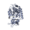
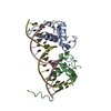
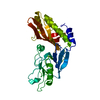
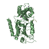
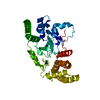
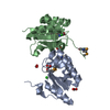
| Title | crystal structure of the streptococcal collagen-like protein 2 globular domain from invasive M3-type group A Streptococcus | ||||||
 Components Components | Collagen-like protein SclB | ||||||
 Keywords Keywords |  STRUCTURAL PROTEIN / six-helix bundle STRUCTURAL PROTEIN / six-helix bundle | ||||||
| Function / homology |  Function and homology information Function and homology informationSingle alpha-helices involved in coiled-coils or other helix-helix interfaces - #2770 / : / M protein-type anchor domain / Collagen triple helix repeat / Collagen triple helix repeat (20 copies) / LPXTG cell wall anchor motif / Single alpha-helices involved in coiled-coils or other helix-helix interfaces / Helix non-globular / Special Similarity search - Domain/homology | ||||||
| Biological species |  Streptococcus pyogenes (bacteria) Streptococcus pyogenes (bacteria) | ||||||
| Method | X-RAY DIFFRACTION / SAD / Resolution: 1.6 Å | ||||||
 Authors Authors | Berisio, R. / Squeglia, F. / Lukomski, S. / Bachert, B. | ||||||
 Citation Citation | Journal: J.Biol.Chem. / Year: 2014 Title: The Crystal Structure of the Streptococcal Collagen-like Protein 2 Globular Domain from Invasive M3-type Group A Streptococcus Shows Significant Similarity to Immunomodulatory HIV Protein gp41. Authors: Squeglia, F. / Bachert, B. / De Simone, A. / Lukomski, S. / Berisio, R. | ||||||
| History |
|
- Structure visualization
| Structure viewer | Molecule: MolmilJmol/JSmol |
|---|
- Downloads & links
-Download
| PDBx/mmCIF format | 4nsm.cif.gz | 47.7 KB | Display | PDBx/mmCIF format |
|---|---|---|---|---|
| PDB format | pdb4nsm.ent.gz | 34.6 KB | Display | PDB format |
| PDBx/mmJSON format | 4nsm.json.gz | Tree view | PDBx/mmJSON format | |
| Others |  Other downloads Other downloads |
-Validation report
| Arichive directory | https://data.pdbj.org/pub/pdb/validation_reports/ns/4nsmftp://data.pdbj.org/pub/pdb/validation_reports/ns/4nsm | HTTPS FTP |
|---|
-Related structure data
| Similar structure data |
|---|


-Links
 PDBj
PDBj- Assembly
| Deposited unit | 
| ||||||||||||
|---|---|---|---|---|---|---|---|---|---|---|---|---|---|
| 1 | 
| ||||||||||||
| Unit cell |
| ||||||||||||
| Components on special symmetry positions |
|
-Components
| #1: Protein | Mass: 10119.227 Da / Num. of mol.: 1 / Fragment: V domain Source method: isolated from a genetically manipulated source Source: (gene. exp.) Streptococcus pyogenes (bacteria) / Strain: ATCC BAA-595 / MGAS315 / Gene: sclB, SpyM3_0738 / Production host: Escherichia coli (E. coli) / References: UniProt: Q8K7M7, UniProt: A0A0H2UUG0*PLUS | ||||
|---|---|---|---|---|---|
| #2: Chemical | Sulfate  Mass: 96.063 Da / Num. of mol.: 2 / Source method: obtained synthetically / Formula: SO4 Mass: 96.063 Da / Num. of mol.: 2 / Source method: obtained synthetically / Formula: SO4#3: Chemical | ChemComp-TRS / | Tris  Mass: 122.143 Da / Num. of mol.: 1 / Source method: obtained synthetically / Formula: C4H12NO3 / Comment: pH buffer*YM Mass: 122.143 Da / Num. of mol.: 1 / Source method: obtained synthetically / Formula: C4H12NO3 / Comment: pH buffer*YM#4: Water | ChemComp-HOH / | Water Mass: 18.015 Da / Num. of mol.: 98 / Source method: isolated from a natural source / Formula: H2O Mass: 18.015 Da / Num. of mol.: 98 / Source method: isolated from a natural source / Formula: H2O |
-Experimental details
-Experiment
| Experiment | Method: X-RAY DIFFRACTION / Number of used crystals: 1 |
|---|
- Sample preparation
| Crystal | Density Matthews: 2.14 Å3/Da / Density % sol: 42.4 % |
|---|---|
| Crystal grow | Temperature: 298 K / Method: evaporation / pH: 6.5 Details: 0.05M Ammonium sulfate, 0.05M BIS-TRIS pH 6.5, 30% v/v Pentaerythritol ethoxylate (15/4 EO/OH), EVAPORATION, temperature 298K |
-Data collection
| Diffraction | Mean temperature: 100 K |
|---|---|
| Diffraction source | Source: ROTATING ANODE / Type: RIGAKU MICROMAX-007 HF / Wavelength: 1.54 Å |
| Detector | Type: RIGAKU SATURN 944 / Detector: CCD / Date: Mar 12, 2013 |
| Radiation | Monochromator: GRAPHITE / Protocol: SINGLE WAVELENGTH / Monochromatic (M) / Laue (L): M / Scattering type: x-ray |
| Radiation wavelength | Wavelength: 1.54 Å / Relative weight: 1 |
| Reflection | Resolution: 1.52→30 Å / Num. all: 13802 / Num. obs: 13802 / % possible obs: 99.2 % / Observed criterion σ(F): 0 / Observed criterion σ(I): 0 / Redundancy: 5.3 % / Rmerge(I) obs: 0.061 |
| Reflection shell | Resolution: 1.52→1.55 Å / Rmerge(I) obs: 0.345 / % possible all: 86.7 |
- Processing
| Software |
| |||||||||||||||||||||||||
|---|---|---|---|---|---|---|---|---|---|---|---|---|---|---|---|---|---|---|---|---|---|---|---|---|---|---|
| Refinement | Method to determine structure: SAD / Resolution: 1.6→14.98 Å / Cor.coef. Fo:Fc: 0.959 / Cor.coef. Fo:Fc free: 0.942 / SU B: 4.632 / SU ML: 0.073 / Cross valid method: THROUGHOUT / ESU R: 0.127 / ESU R Free: 0.1 Stereochemistry target values: MAXIMUM LIKELIHOOD WITH PHASES
| |||||||||||||||||||||||||
| Solvent computation | Ion probe radii: 0.8 Å / Shrinkage radii: 0.8 Å / VDW probe radii: 1.4 Å / Solvent model: BABINET MODEL WITH MASK | |||||||||||||||||||||||||
| Displacement parameters | Biso mean: 20.793 Å2
| |||||||||||||||||||||||||
| Refinement step | Cycle: LAST / Resolution: 1.6→14.98 Å
| |||||||||||||||||||||||||
| Refine LS restraints |
| |||||||||||||||||||||||||
| LS refinement shell | Resolution: 1.6→1.641 Å / Total num. of bins used: 20
|
