[English] 日本語
Yorodumi
- PDB-4nob: Crystal structure of the 1st Ig domain from mouse Polymeric Immun... -

+
Open data


ID or keywords:

Loading...

-
Basic information

Entry
Database: PDB / ID: 4nob
Title
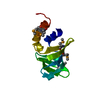
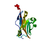
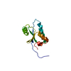
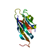
Crystal structure of the 1st Ig domain from mouse Polymeric Immunoglobulin receptor [PSI-NYSGRC-006220]
ComponentsPolymeric immunoglobulin receptor
KeywordsIMMUNE SYSTEM / ortholog / Ig domain / Structural genomics / PSI-Biology / New York Structural Genomics Research Consortium / NYSGRC / Immune Function Network / IFN / Atoms-to-Animals: The Immune Function Network
Function / homology
Function and homology information


polymeric immunoglobulin receptor activity / immunoglobulin transcytosis in epithelial cells mediated by polymeric immunoglobulin receptor / polymeric immunoglobulin binding / secretory IgA immunoglobulin complex / Fc receptor signaling pathway / detection of chemical stimulus involved in sensory perception of bitter taste / epidermal growth factor receptor binding / receptor clustering / Neutrophil degranulation / epidermal growth factor receptor signaling pathway ...polymeric immunoglobulin receptor activity / immunoglobulin transcytosis in epithelial cells mediated by polymeric immunoglobulin receptor / polymeric immunoglobulin binding / secretory IgA immunoglobulin complex / Fc receptor signaling pathway / detection of chemical stimulus involved in sensory perception of bitter taste / epidermal growth factor receptor binding / receptor clustering / Neutrophil degranulation / epidermal growth factor receptor signaling pathway / recycling endosome membrane / receptor complex / extracellular space / plasma membrane
Similarity search - Function
Immunoglobulin V-Type / Immunoglobulin V-set domain / Immunoglobulin V-set domain / Immunoglobulin subtype / Immunoglobulin / Ig-like domain profile. / Immunoglobulin-like domain / Immunoglobulin-like domain superfamily / Immunoglobulins / Immunoglobulin-like fold ...Immunoglobulin V-Type / Immunoglobulin V-set domain / Immunoglobulin V-set domain / Immunoglobulin subtype / Immunoglobulin / Ig-like domain profile. / Immunoglobulin-like domain / Immunoglobulin-like domain superfamily / Immunoglobulins / Immunoglobulin-like fold / Immunoglobulin-like / Sandwich / Mainly Beta
Similarity search - Domain/homology
Polymeric immunoglobulin receptor
Similarity search - Component
Biological speciesMus musculus (house mouse)
MethodX-RAY DIFFRACTION / SYNCHROTRON / MOLECULAR REPLACEMENT / Resolution: 1.51 Å
AuthorsKumar, P.R. / Banu, R. / Bhosle, R. / Calarese, D.A. / Celikgil, A. / Chamala, S. / Chan, M.K. / Chowdhury, S. / Fiser, A. / Garforth, S.J. ...Kumar, P.R. / Banu, R. / Bhosle, R. / Calarese, D.A. / Celikgil, A. / Chamala, S. / Chan, M.K. / Chowdhury, S. / Fiser, A. / Garforth, S.J. / Glenn, A.S. / Hillerich, B. / Khafizov, K. / Attonito, J. / Love, J.D. / Patel, H. / Patel, R. / Seidel, R.D. / Smith, B. / Stead, M. / Casadevall, A. / Almo, S.C. / New York Structural Genomics Research Consortium (NYSGRC) / Atoms-to-Animals: The Immune Function Network (IFN)
CitationJournal: to be published
Title: Crystal structure of the 1st Ig domain from mouse Polymeric Immunoglobulin receptor
Authors: Kumar, P.R. / Casadevall, A. / Almo, S.C.
History
DepositionNov 19, 2013Deposition site: RCSB / Processing site: RCSB
Revision 1.0Dec 4, 2013Provider: repository / Type: Initial release
Revision 1.1Jul 29, 2020Group: Data collection / Database references ...Data collection / Database references / Derived calculations / Structure summary
Category: chem_comp / entity ...chem_comp / entity / pdbx_chem_comp_identifier / pdbx_entity_nonpoly / pdbx_struct_conn_angle / struct_conn / struct_ref_seq_dif / struct_site / struct_site_gen
Item: _chem_comp.name / _chem_comp.type ..._chem_comp.name / _chem_comp.type / _entity.pdbx_description / _pdbx_entity_nonpoly.name / _pdbx_struct_conn_angle.ptnr1_auth_comp_id / _pdbx_struct_conn_angle.ptnr1_auth_seq_id / _pdbx_struct_conn_angle.ptnr1_label_asym_id / _pdbx_struct_conn_angle.ptnr1_label_atom_id / _pdbx_struct_conn_angle.ptnr1_label_comp_id / _pdbx_struct_conn_angle.ptnr1_label_seq_id / _pdbx_struct_conn_angle.ptnr3_auth_comp_id / _pdbx_struct_conn_angle.ptnr3_auth_seq_id / _pdbx_struct_conn_angle.ptnr3_label_asym_id / _pdbx_struct_conn_angle.ptnr3_label_atom_id / _pdbx_struct_conn_angle.ptnr3_label_comp_id / _pdbx_struct_conn_angle.ptnr3_label_seq_id / _pdbx_struct_conn_angle.value / _struct_conn.pdbx_dist_value / _struct_conn.pdbx_leaving_atom_flag / _struct_conn.pdbx_role / _struct_conn.ptnr1_auth_comp_id / _struct_conn.ptnr1_auth_seq_id / _struct_conn.ptnr1_label_asym_id / _struct_conn.ptnr1_label_atom_id / _struct_conn.ptnr1_label_comp_id / _struct_conn.ptnr1_label_seq_id / _struct_conn.ptnr2_auth_comp_id / _struct_conn.ptnr2_auth_seq_id / _struct_conn.ptnr2_label_asym_id / _struct_conn.ptnr2_label_atom_id / _struct_conn.ptnr2_label_comp_id / _struct_ref_seq_dif.details
Description: Carbohydrate remediation / Provider: repository / Type: Remediation
Revision 1.2Sep 20, 2023Group: Data collection / Database references ...Data collection / Database references / Refinement description / Structure summary
Category: chem_comp / chem_comp_atom ...chem_comp / chem_comp_atom / chem_comp_bond / database_2 / pdbx_initial_refinement_model
Item: _chem_comp.pdbx_synonyms / _database_2.pdbx_DOI / _database_2.pdbx_database_accession

-
Structure visualization

Structure viewerMolecule:
MolmilJmol/JSmol

Downloads & links

-
Assembly

Deposited unit
A: Polymeric immunoglobulin receptor
hetero molecules


Theoretical massNumber of molelcules
Total (without water)14,3716
Polymers13,9391
Non-polymers4325
Water1,982110
1


  • Idetical with deposited unit
  • defined by author&software
TypeNameSymmetry operationNumber
identity operation1_555x,y,z1
MethodPISA
Unit cell
Length a, b, c (Å)40.275, 46.803, 62.062
Angle α, β, γ (deg.)90.000, 90.000, 90.000
Int Tables number19
Space group name H-MP212121

-
Components

#1: Protein Polymeric immunoglobulin receptor / / PIgR / Poly-Ig receptor / Secretory component


Mass: 13939.219 Da / Num. of mol.: 1 / Fragment: Ig-like V-type 1 domain residues 20-133
Source method: isolated from a genetically manipulated source
Source: (gene. exp.) Mus musculus (house mouse) / Gene: Pigr / Plasmid: pIEX / Production host: Trichoplusia ni (cabbage looper) / Strain (production host): Hi5 / References: UniProt: O70570
#2: Sugar ChemComp-NAG / 2-acetamido-2-deoxy-beta-D-glucopyranose / N-acetyl-beta-D-glucosamine / 2-acetamido-2-deoxy-beta-D-glucose / 2-acetamido-2-deoxy-D-glucose / 2-acetamido-2-deoxy-glucose / N-ACETYL-D-GLUCOSAMINE / N-Acetylglucosamine


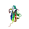
Type: D-saccharide, beta linking / Mass: 221.208 Da / Num. of mol.: 1
Source method: isolated from a genetically manipulated source
Formula: C8H15NO6
IdentifierTypeProgram
DGlcpNAcbCONDENSED IUPAC CARBOHYDRATE SYMBOLGMML 1.0
N-acetyl-b-D-glucopyranosamineCOMMON NAMEGMML 1.0
b-D-GlcpNAcIUPAC CARBOHYDRATE SYMBOLPDB-CARE 1.0
GlcNAcSNFG CARBOHYDRATE SYMBOLGMML 1.0
#3: Chemical ChemComp-MG / MAGNESIUM ION


Mass: 24.305 Da / Num. of mol.: 1 / Source method: obtained synthetically / Formula: Mg
#4: Chemical ChemComp-EDO / 1,2-ETHANEDIOL / ETHYLENE GLYCOL / Ethylene glycol


Mass: 62.068 Da / Num. of mol.: 3 / Source method: obtained synthetically / Formula: C2H6O2
#5: Water ChemComp-HOH / water / Water


Mass: 18.015 Da / Num. of mol.: 110 / Source method: isolated from a natural source / Formula: H2O

-
Experimental details

-
Experiment

ExperimentMethod: X-RAY DIFFRACTION / Number of used crystals: 1

-
Sample preparation

CrystalDensity Matthews: 2.1 Å3/Da / Density % sol: 41.38 %
Crystal growTemperature: 298 K / Method: vapor diffusion, sitting drop / pH: 5.5
Details: Protein (20 mM Hepes, pH 7.5, 150 mM NaCl, 5% glycerol), Reservoir (0.2M MgCl2, 0.1M Bis-Tris-HCl, 25% (v/v) PEG 3350), Cryoprotection (33% Ethylene glycol), Vapor Diffusion, Sitting Drop, temperature 298K

-
Data collection

DiffractionMean temperature: 100 K
Diffraction sourceSource: SYNCHROTRON / Site: NSLS / Beamline: X25 / Wavelength: 1.075 Å
DetectorType: DECTRIS PILATUS 6M / Detector: PIXEL / Date: Oct 24, 2013 / Details: MIRRORS
RadiationMonochromator: Si(111) / Protocol: SINGLE WAVELENGTH / Monochromatic (M) / Laue (L): M / Scattering type: x-ray
Radiation wavelengthWavelength: 1.075 Å / Relative weight: 1
ReflectionResolution: 1.51→50 Å / Num. obs: 18980 / % possible obs: 99.4 % / Redundancy: 11.9 % / Rmerge(I) obs: 0.077 / Χ2: 0.974 / Net I/σ(I): 8.1
Reflection shell
Resolution (Å)Redundancy (%)Rmerge(I) obsNum. unique allΧ2Diffraction-ID% possible all
1.51-1.5480.3538531.043193.3
1.54-1.569.40.339231.058197.8
1.56-1.5910.80.3079301.0831100
1.59-1.6312.20.2919321.043198.6
1.63-1.6612.30.2519161.0871100
1.66-1.7120.239421.046199.3
1.7-1.7412.50.2119441.0971100
1.74-1.7912.50.1879281.118199.5
1.79-1.8412.20.1619501.119199.8
1.84-1.912.70.1419431.0781100
1.9-1.9712.10.1189341.052199.8
1.97-2.0512.20.1029521.0261100
2.05-2.1412.30.0949580.9891100
2.14-2.2612.50.0829500.911100
2.26-2.412.60.0779640.8671100
2.4-2.5812.40.079550.769199.9
2.58-2.8412.40.0639680.7261100
2.84-3.25130.0599760.6951100
3.25-4.112.30.0599950.7341100
4.1-5011.50.06810671.069199.9

-
Processing

Software
NameVersionClassificationNB
SCALEPACKdata scaling
REFMACrefinement
PDB_EXTRACT3.11data extraction
CBASSdata collection
HKL-3000data reduction
MOLREPphasing
RefinementMethod to determine structure: MOLECULAR REPLACEMENT
Starting model: 1XED
Resolution: 1.51→37.37 Å / Cor.coef. Fo:Fc: 0.965 / Cor.coef. Fo:Fc free: 0.957 / WRfactor Rfree: 0.2109 / WRfactor Rwork: 0.1745 / Occupancy max: 1 / Occupancy min: 0.5 / FOM work R set: 0.8837 / SU B: 1.059 / SU ML: 0.04 / SU R Cruickshank DPI: 0.0684 / SU Rfree: 0.0704 / Cross valid method: THROUGHOUT / σ(F): 0 / ESU R: 0.068 / ESU R Free: 0.07 / Stereochemistry target values: MAXIMUM LIKELIHOOD
Details: HYDROGENS HAVE BEEN ADDED IN THE RIDING POSITIONS U VALUES : REFINED INDIVIDUALLY
RfactorNum. reflection% reflectionSelection details
Rfree0.1891 945 5.1 %RANDOM
Rwork0.161 ---
obs0.1625 18374 96.48 %-
Solvent computationIon probe radii: 0.8 Å / Shrinkage radii: 0.8 Å / VDW probe radii: 1.2 Å / Solvent model: MASK
Displacement parametersBiso max: 82.96 Å2 / Biso mean: 17.001 Å2 / Biso min: 8.44 Å2
Baniso -1Baniso -2Baniso -3
1--0.24 Å2-0 Å20 Å2
2---0.32 Å20 Å2
3---0.56 Å2
Refinement stepCycle: LAST / Resolution: 1.51→37.37 Å
ProteinNucleic acidLigandSolventTotal
Num. atoms858 0 27 110 995
Refine LS restraints
Refine-IDTypeDev idealDev ideal targetNumber
X-RAY DIFFRACTIONr_bond_refined_d0.0230.02907
X-RAY DIFFRACTIONr_bond_other_d0.0010.02817
X-RAY DIFFRACTIONr_angle_refined_deg2.2141.9771227
X-RAY DIFFRACTIONr_angle_other_deg0.9473.0041880
X-RAY DIFFRACTIONr_dihedral_angle_1_deg5.6855112
X-RAY DIFFRACTIONr_dihedral_angle_2_deg36.6522540
X-RAY DIFFRACTIONr_dihedral_angle_3_deg12.29515141
X-RAY DIFFRACTIONr_dihedral_angle_4_deg16.227154
X-RAY DIFFRACTIONr_chiral_restr0.1380.2137
X-RAY DIFFRACTIONr_gen_planes_refined0.0120.0211029
X-RAY DIFFRACTIONr_gen_planes_other0.0010.02201
X-RAY DIFFRACTIONr_mcbond_it1.9331.326451
X-RAY DIFFRACTIONr_mcbond_other1.931.321450
X-RAY DIFFRACTIONr_mcangle_it3.1841.968562
LS refinement shellResolution: 1.509→1.548 Å / Total num. of bins used: 20
RfactorNum. reflection% reflection
Rfree0.227 39 -
Rwork0.171 968 -
all-1007 -
obs--73.66 %

+
About Yorodumi

-
News

-
Feb 9, 2022. New format data for meta-information of EMDB entries

New format data for meta-information of EMDB entries

  • Version 3 of the EMDB header file is now the official format.
  • The previous official version 1.9 will be removed from the archive.

Related info.:EMDB header

External links:wwPDB to switch to version 3 of the EMDB data model

-
Aug 12, 2020. Covid-19 info

Covid-19 info

URL: https://pdbj.org/emnavi/covid19.php

New page: Covid-19 featured information page in EM Navigator.

Related info.:Covid-19 info / Mar 5, 2020. Novel coronavirus structure data

+
Mar 5, 2020. Novel coronavirus structure data

Novel coronavirus structure data

Related info.:Yorodumi Speices / Aug 12, 2020. Covid-19 info

External links:COVID-19 featured content - PDBj / Molecule of the Month (242):Coronavirus Proteases

+
Jan 31, 2019. EMDB accession codes are about to change! (news from PDBe EMDB page)

EMDB accession codes are about to change! (news from PDBe EMDB page)

  • The allocation of 4 digits for EMDB accession codes will soon come to an end. Whilst these codes will remain in use, new EMDB accession codes will include an additional digit and will expand incrementally as the available range of codes is exhausted. The current 4-digit format prefixed with “EMD-” (i.e. EMD-XXXX) will advance to a 5-digit format (i.e. EMD-XXXXX), and so on. It is currently estimated that the 4-digit codes will be depleted around Spring 2019, at which point the 5-digit format will come into force.
  • The EM Navigator/Yorodumi systems omit the EMD- prefix.

Related info.:Q: What is EMD? / ID/Accession-code notation in Yorodumi/EM Navigator

External links:EMDB Accession Codes are Changing Soon! / Contact to PDBj

+
Jul 12, 2017. Major update of PDB

Major update of PDB

  • wwPDB released updated PDB data conforming to the new PDBx/mmCIF dictionary.
  • This is a major update changing the version number from 4 to 5, and with Remediation, in which all the entries are updated.
  • In this update, many items about electron microscopy experimental information are reorganized (e.g. em_software).
  • Now, EM Navigator and Yorodumi are based on the updated data.

External links:wwPDB Remediation / Enriched Model Files Conforming to OneDep Data Standards Now Available in the PDB FTP Archive

-
Yorodumi

Thousand views of thousand structures

  • Yorodumi is a browser for structure data from EMDB, PDB, SASBDB, etc.
  • This page is also the successor to EM Navigator detail page, and also detail information page/front-end page for Omokage search.
  • The word "yorodu" (or yorozu) is an old Japanese word meaning "ten thousand". "mi" (miru) is to see.

Related info.:EMDB / PDB / SASBDB / Comparison of 3 databanks / Yorodumi Search / Aug 31, 2016. New EM Navigator & Yorodumi / Yorodumi Papers / Jmol/JSmol / Function and homology information / Changes in new EM Navigator and Yorodumi

Read more