 Movie
Movie Controller


+ Open data

- Basic information
| Entry | Database: PDB / ID: 4noa | ||||||
|---|---|---|---|---|---|---|---|
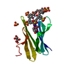
| Title | Truncated minor pilin PilE from Pseudomonas aeruginosa | ||||||
 Components Components | Type 4 fimbrial biogenesis protein PilE | ||||||
 Keywords Keywords | UNKNOWN FUNCTION | ||||||
| Function / homology |  Function and homology information Function and homology information type IV pilus assembly / type IV pilus-dependent motility / pilus assembly / membrane => GO:0016020 / plasma membrane type IV pilus assembly / type IV pilus-dependent motility / pilus assembly / membrane => GO:0016020 / plasma membraneSimilarity search - Function | ||||||
| Biological species |   Pseudomonas aeruginosa (bacteria) Pseudomonas aeruginosa (bacteria) | ||||||
| Method | X-RAY DIFFRACTION / SYNCHROTRON / SAD / Resolution: 1.25 Å | ||||||
 Authors Authors | Nguyen, Y. / Sugiman-Marangos, S.N. / Bell, S. / Junop, M.S. / Burrows, L.L. | ||||||
 Citation Citation | Journal: To be Published Title: Crystal structure of truncated minor pilin PilE from Pseudomonas aeruginosa Authors: Nguyen, Y. / Sugiman-Marangos, S.N. / Bell, S. / Harvey, H. / Junop, M.S. / Burrows, L.L. | ||||||
| History |
|
- Structure visualization
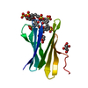
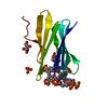
| Structure viewer | Molecule: MolmilJmol/JSmol |
|---|
- Downloads & links
-Download
| PDBx/mmCIF format | 4noa.cif.gz | 34.8 KB | Display | PDBx/mmCIF format |
|---|---|---|---|---|
| PDB format | pdb4noa.ent.gz | 26.4 KB | Display | PDB format |
| PDBx/mmJSON format | 4noa.json.gz | Tree view | PDBx/mmJSON format | |
| Others |  Other downloads Other downloads |
-Validation report
| Arichive directory | https://data.pdbj.org/pub/pdb/validation_reports/no/4noaftp://data.pdbj.org/pub/pdb/validation_reports/no/4noa | HTTPS FTP |
|---|
-Related structure data
| Similar structure data |
|---|


-Links
 PDBj
PDBj
- Assembly
| Deposited unit | 
| ||||||||
|---|---|---|---|---|---|---|---|---|---|
| 1 |
| ||||||||
| Unit cell |
| ||||||||
| Details | AUTHORS STATE THAT THE BIOLOGICAL ASSEMBLY IN UNKNOWN. |
-Components
| #1: Protein | Mass: 12093.130 Da / Num. of mol.: 1 / Fragment: UNP residues 36-141 Source method: isolated from a genetically manipulated source Source: (gene. exp.) Pseudomonas aeruginosa (bacteria) / Strain: ATCC 15692 / PAO1 / 1C / PRS 101 / LMG 12228 / Gene: PA4556, pilE / Plasmid: pET151-D-topo / Production host: Escherichia coli (E. coli) / Strain (production host): Origami B(DE3) / References: UniProt: G3XD43 |
|---|---|
| #2: Water | ChemComp-HOH / Water Mass: 18.015 Da / Num. of mol.: 244 / Source method: isolated from a natural source / Formula: H2O Mass: 18.015 Da / Num. of mol.: 244 / Source method: isolated from a natural source / Formula: H2O |
-Experimental details
-Experiment
| Experiment | Method: X-RAY DIFFRACTION / Number of used crystals: 1 |
|---|
- Sample preparation
| Crystal | Density Matthews: 2.42 Å3/Da / Density % sol: 49.12 % |
|---|---|
| Crystal grow | Temperature: 293 K / Method: vapor diffusion, hanging drop / pH: 8 Details: 0.2 M Ammonium tartrate dibasic, 20% (w/v) PEG 3350, pH 8.0, VAPOR DIFFUSION, HANGING DROP, temperature 293K |
-Data collection
| Diffraction | Mean temperature: 100 K |
|---|---|
| Diffraction source | Source: SYNCHROTRON / Site: NSLS  / Beamline: X25 / Wavelength: 0.979 Å / Beamline: X25 / Wavelength: 0.979 Å |
| Detector | Type: PSI PILATUS 6M / Detector: PIXEL / Date: Sep 25, 2012 / Details: Mirrors |
| Radiation | Monochromator: Si(111) / Protocol: SINGLE WAVELENGTH / Monochromatic (M) / Laue (L): M / Scattering type: x-ray |
| Radiation wavelength | Wavelength: 0.979 Å / Relative weight: 1 |
| Reflection | Resolution: 1.25→50 Å / Num. all: 30749 / Num. obs: 30749 / % possible obs: 95.4 % / Observed criterion σ(F): 0 / Observed criterion σ(I): 0 / Redundancy: 6.4 % / Biso Wilson estimate: 8.28 Å2 / Rmerge(I) obs: 0.084 / Rsym value: 0.084 / Net I/σ(I): 20.7 |
| Reflection shell | Resolution: 1.25→1.27 Å / Redundancy: 4.5 % / Rmerge(I) obs: 0.21 / Mean I/σ(I) obs: 7.6 / Num. unique all: 1462 / Rsym value: 0.21 / % possible all: 89.6 |
- Processing
| Software |
| |||||||||||||||||||||||||||||||||||||||||||||||||||||||||||||||||||||||||||||||||||||||||||||||||||||||||
|---|---|---|---|---|---|---|---|---|---|---|---|---|---|---|---|---|---|---|---|---|---|---|---|---|---|---|---|---|---|---|---|---|---|---|---|---|---|---|---|---|---|---|---|---|---|---|---|---|---|---|---|---|---|---|---|---|---|---|---|---|---|---|---|---|---|---|---|---|---|---|---|---|---|---|---|---|---|---|---|---|---|---|---|---|---|---|---|---|---|---|---|---|---|---|---|---|---|---|---|---|---|---|---|---|---|---|
| Refinement | Method to determine structure: SAD / Resolution: 1.25→43.181 Å / SU ML: 0.12 / σ(F): 0 / Phase error: 14.68 / Stereochemistry target values: MLHL
| |||||||||||||||||||||||||||||||||||||||||||||||||||||||||||||||||||||||||||||||||||||||||||||||||||||||||
| Solvent computation | Shrinkage radii: 0.6 Å / VDW probe radii: 0.9 Å / Solvent model: FLAT BULK SOLVENT MODEL / Bsol: 43.969 Å2 / ksol: 0.442 e/Å3 | |||||||||||||||||||||||||||||||||||||||||||||||||||||||||||||||||||||||||||||||||||||||||||||||||||||||||
| Displacement parameters | Biso mean: 11.51 Å2
| |||||||||||||||||||||||||||||||||||||||||||||||||||||||||||||||||||||||||||||||||||||||||||||||||||||||||
| Refinement step | Cycle: LAST / Resolution: 1.25→43.181 Å
| |||||||||||||||||||||||||||||||||||||||||||||||||||||||||||||||||||||||||||||||||||||||||||||||||||||||||
| Refine LS restraints |
| |||||||||||||||||||||||||||||||||||||||||||||||||||||||||||||||||||||||||||||||||||||||||||||||||||||||||
| LS refinement shell |
|
