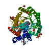
| Deposited unit | A: Complement C1q tumor necrosis factor-related protein 5
B: Complement C1q tumor necrosis factor-related protein 5
C: Complement C1q tumor necrosis factor-related protein 5
hetero molecules
| Theoretical mass | Number of molelcules |
|---|
| Total (without water) | 50,796 | 26 |
|---|
| Polymers | 49,273 | 3 |
|---|
| Non-polymers | 1,524 | 23 |
|---|
| Water | 5,296 | 294 |
|---|
|
|---|
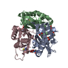
| 1 | A: Complement C1q tumor necrosis factor-related protein 5
hetero molecules
A: Complement C1q tumor necrosis factor-related protein 5
hetero molecules
A: Complement C1q tumor necrosis factor-related protein 5
hetero molecules
| Theoretical mass | Number of molelcules |
|---|
| Total (without water) | 51,973 | 45 |
|---|
| Polymers | 49,273 | 3 |
|---|
| Non-polymers | 2,700 | 42 |
|---|
| Water | 54 | 3 |
|---|
| Type | Name | Symmetry operation | Number |
|---|
| identity operation | 1_555 | x,y,z | 1 | | crystal symmetry operation | 2_555 | -y,x-y,z | 1 | | crystal symmetry operation | 3_555 | -x+y,-x,z | 1 |
| Buried area | 12630 Å2 |
|---|
| ΔGint | -3 kcal/mol |
|---|
| Surface area | 15660 Å2 |
|---|
| Method | PISA |
|---|
|
|---|
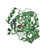
| 2 | B: Complement C1q tumor necrosis factor-related protein 5
hetero molecules
B: Complement C1q tumor necrosis factor-related protein 5
hetero molecules
B: Complement C1q tumor necrosis factor-related protein 5
hetero molecules
| Theoretical mass | Number of molelcules |
|---|
| Total (without water) | 50,957 | 27 |
|---|
| Polymers | 49,273 | 3 |
|---|
| Non-polymers | 1,685 | 24 |
|---|
| Water | 54 | 3 |
|---|
| Type | Name | Symmetry operation | Number |
|---|
| identity operation | 1_555 | x,y,z | 1 | | crystal symmetry operation | 2_565 | -y,x-y+1,z | 1 | | crystal symmetry operation | 3_455 | -x+y-1,-x,z | 1 |
| Buried area | 9780 Å2 |
|---|
| ΔGint | -42 kcal/mol |
|---|
| Surface area | 16280 Å2 |
|---|
| Method | PISA |
|---|
|
|---|
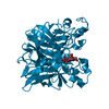
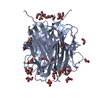
| 3 | C: Complement C1q tumor necrosis factor-related protein 5
hetero molecules
C: Complement C1q tumor necrosis factor-related protein 5
hetero molecules
C: Complement C1q tumor necrosis factor-related protein 5
hetero molecules
| Theoretical mass | Number of molelcules |
|---|
| Total (without water) | 49,459 | 6 |
|---|
| Polymers | 49,273 | 3 |
|---|
| Non-polymers | 186 | 3 |
|---|
| Water | 54 | 3 |
|---|
| Type | Name | Symmetry operation | Number |
|---|
| identity operation | 1_555 | x,y,z | 1 | | crystal symmetry operation | 2_555 | -y,x-y,z | 1 | | crystal symmetry operation | 3_555 | -x+y,-x,z | 1 |
| Buried area | 7830 Å2 |
|---|
| ΔGint | -8 kcal/mol |
|---|
| Surface area | 15600 Å2 |
|---|
| Method | PISA |
|---|
|
|---|
| Unit cell | | Length a, b, c (Å) | 50.603, 50.603, 268.531 |
|---|
| Angle α, β, γ (deg.) | 90.00, 90.00, 120.00 |
|---|
| Int Tables number | 173 |
|---|
| Space group name H-M | P63 |
|---|
|
|---|
| Components on special symmetry positions | | ID | Model | Components |
|---|
| 1 | 1 | A-313- SO4 | | 2 | 1 | A-313-SO4 | | 3 | 1 | B-306-SO4 | | 4 | 1 | B-306-SO4 | | 5 | 1 | A-401- HOH | | 6 | 1 | A-429-HOH | | 7 | 1 | A-510-HOH | | 8 | 1 | A-514-HOH | | 9 | 1 | A-518-HOH | | 10 | 1 | B-401-HOH | | 11 | 1 | B-427-HOH | | 12 | 1 | B-503-HOH | | 13 | 1 | B-509-HOH | | 14 | 1 | C-401-HOH |
|
|---|
| Noncrystallographic symmetry (NCS) | NCS oper: | ID | Code | Matrix | Vector |
|---|
| 1 | given(1), (1), (1) | | 2 | given(-0.496073, 0.868275, -0.003155), (0.868273, 0.496081, 0.002471), (0.00371, -0.001514, -0.999992)| -25.2918, 14.73086, -11.95059 | | | | |
|
|---|
 Movie
Movie Controller

 Yorodumi
 Open data
 Basic information
 Components
 Keywords
 Function and homology information
 Authors
 Citation
 Structure visualization
 Downloads & links
 Other downloads
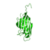


 PDBj
PDBj Assembly
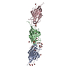


 Mass: 62.068 Da / Num. of mol.: 18 / Source method: obtained synthetically / Formula: C2H6O2
 Mass: 96.063 Da / Num. of mol.: 3 / Source method: obtained synthetically / Formula: SO4
 Mass: 59.044 Da / Num. of mol.: 2 / Source method: obtained synthetically / Formula: C2H3O2
 Sample preparation
 / Beamline: 24-ID-C / Wavelength: 0.9792 Å
 Processing