 Movie
Movie Controller

[English] 日本語
 Yorodumi
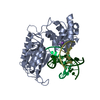
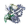
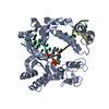
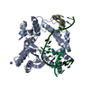
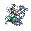
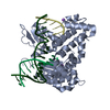
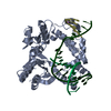
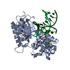
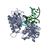
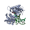
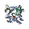
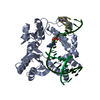
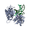
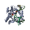
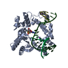
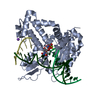
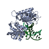
- PDB-4nlz: Structure of human DNA polymerase beta complexed with nicked DNA ... -
+ Open data

- Basic information
| Entry | Database: PDB / ID: 4nlz | ||||||
|---|---|---|---|---|---|---|---|
| Title | Structure of human DNA polymerase beta complexed with nicked DNA containing a mismatched template 8BrG and incoming GTP | ||||||
 Components Components |
| ||||||
 Keywords Keywords |  TRANSFERASE / LYASE/DNA / DNA binding / helix-turn-helix motif / polymerase fold / nucleotidyl transferase / nucleus / LYASE-DNA complex TRANSFERASE / LYASE/DNA / DNA binding / helix-turn-helix motif / polymerase fold / nucleotidyl transferase / nucleus / LYASE-DNA complex | ||||||
| Function / homology |  Function and homology information Function and homology informationResolution of AP sites via the single-nucleotide replacement pathway / immunoglobulin heavy chain V-D-J recombination / Resolution of AP sites via the multiple-nucleotide patch replacement pathway / Abasic sugar-phosphate removal via the single-nucleotide replacement pathway / APEX1-Independent Resolution of AP Sites via the Single Nucleotide Replacement Pathway / Lyases; Carbon-oxygen lyases; Other carbon-oxygen lyases / homeostasis of number of cells / 5'-deoxyribose-5-phosphate lyase activity / POLB-Dependent Long Patch Base Excision Repair / PCNA-Dependent Long Patch Base Excision Repair ...Resolution of AP sites via the single-nucleotide replacement pathway / immunoglobulin heavy chain V-D-J recombination / Resolution of AP sites via the multiple-nucleotide patch replacement pathway / Abasic sugar-phosphate removal via the single-nucleotide replacement pathway / APEX1-Independent Resolution of AP Sites via the Single Nucleotide Replacement Pathway / Lyases; Carbon-oxygen lyases; Other carbon-oxygen lyases / homeostasis of number of cells / 5'-deoxyribose-5-phosphate lyase activity / POLB-Dependent Long Patch Base Excision Repair / PCNA-Dependent Long Patch Base Excision Repair / pyrimidine dimer repair / response to hyperoxia / somatic hypermutation of immunoglobulin genes / lymph node development / salivary gland morphogenesis / spleen development / DNA-(apurinic or apyrimidinic site) endonuclease activity / class I DNA-(apurinic or apyrimidinic site) endonuclease activity / DNA-(apurinic or apyrimidinic site) lyase / base-excision repair, gap-filling / response to gamma radiation / base-excision repair / spindle microtubule / DNA-templated DNA replication / double-strand break repair via nonhomologous end joining / intrinsic apoptotic signaling pathway in response to DNA damage / microtubule binding / neuron apoptotic process / response to ethanol / microtubule / in utero embryonic development / damaged DNA binding / DNA-directed DNA polymerase / DNA-directed DNA polymerase activity / lyase activity / Ub-specific processing proteases / inflammatory response / DNA repair / DNA damage response / enzyme binding / protein-containing complex / nucleoplasm / metal ion binding / nucleus / cytoplasmSimilarity search - Function | ||||||
| Biological species |  Homo sapiens (human) Homo sapiens (human) | ||||||
| Method | X-RAY DIFFRACTION / MOLECULAR REPLACEMENT / Resolution: 2.683 Å | ||||||
 Authors Authors | Koag, M.-C. / Min, K. / Monzingo, A.F. / Lee, S. | ||||||
 Citation Citation | Journal: J.Biol.Chem. / Year: 2014 Title: Structural basis for promutagenicity of 8-halogenated Guanine. Authors: Koag, M.C. / Min, K. / Lee, S. | ||||||
| History |
|
- Structure visualization
| Structure viewer | Molecule: MolmilJmol/JSmol |
|---|
- Downloads & links
-Download
| PDBx/mmCIF format | 4nlz.cif.gz | 99.5 KB | Display | PDBx/mmCIF format |
|---|---|---|---|---|
| PDB format | pdb4nlz.ent.gz | 71.1 KB | Display | PDB format |
| PDBx/mmJSON format | 4nlz.json.gz | Tree view | PDBx/mmJSON format | |
| Others |  Other downloads Other downloads |
-Validation report
| Arichive directory | https://data.pdbj.org/pub/pdb/validation_reports/nl/4nlzftp://data.pdbj.org/pub/pdb/validation_reports/nl/4nlz | HTTPS FTP |
|---|
-Related structure data
| Related structure data |  4m2yC  4m47C  4nlkC 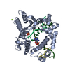 4nlnC 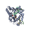 4nm1C  4nm2C C: citing same article ( |
|---|---|
| Similar structure data |


-Links
 PDBj
PDBj


- Assembly
| Deposited unit | 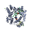
| ||||||||
|---|---|---|---|---|---|---|---|---|---|
| 1 |
| ||||||||
| Unit cell |
|
-Components
-Protein , 1 types, 1 molecules A
| #1: Protein | Mass: 37536.770 Da / Num. of mol.: 1 Source method: isolated from a genetically manipulated source Source: (gene. exp.) Homo sapiens (human) / Gene: POLB / Production host:  Escherichia coli (E. coli) Escherichia coli (E. coli)References: UniProt: P06746, DNA-directed DNA polymerase, Lyases; Carbon-oxygen lyases; Other carbon-oxygen lyases |
|---|
-DNA chain , 3 types, 3 molecules TPD
| #2: DNA chain | Mass: 4923.040 Da / Num. of mol.: 1 / Source method: obtained synthetically / Details: template |
|---|---|
| #3: DNA chain | Mass: 3414.234 Da / Num. of mol.: 1 / Source method: obtained synthetically / Details: up primer |
| #4: DNA chain | Mass: 1536.035 Da / Num. of mol.: 1 / Source method: obtained synthetically / Details: down primer |
-Non-polymers , 4 types, 61 molecules 


| #5: Chemical | ChemComp-MG /  Mass: 24.305 Da / Num. of mol.: 1 / Source method: obtained synthetically / Formula: Mg Mass: 24.305 Da / Num. of mol.: 1 / Source method: obtained synthetically / Formula: Mg | ||||
|---|---|---|---|---|---|
| #6: Chemical |  Mass: 22.990 Da / Num. of mol.: 2 / Source method: obtained synthetically / Formula: Na Mass: 22.990 Da / Num. of mol.: 2 / Source method: obtained synthetically / Formula: Na#7: Chemical | ChemComp-PO4 / | Phosphate Mass: 94.971 Da / Num. of mol.: 1 / Source method: obtained synthetically / Formula: PO4 Mass: 94.971 Da / Num. of mol.: 1 / Source method: obtained synthetically / Formula: PO4#8: Water | ChemComp-HOH / | WaterMass: 18.015 Da / Num. of mol.: 57 / Source method: isolated from a natural source / Formula: H2O |
-Experimental details
-Experiment
| Experiment | Method: X-RAY DIFFRACTION / Number of used crystals: 1 |
|---|
- Sample preparation
| Crystal | Density Matthews: 2.41 Å3/Da / Density % sol: 48.93 % |
|---|---|
| Crystal grow | Method: vapor diffusion, sitting drop / pH: 7.5 Details: 14-23% PEG3400, 350 mM sodium acetate, 50 mM imidazole, pH 7.5, VAPOR DIFFUSION, SITTING DROP |
-Data collection
| Diffraction | Mean temperature: 100 K |
|---|---|
| Diffraction source | Source: ROTATING ANODE / Type: RIGAKU MICROMAX-007 HF / Wavelength: 1.5418 Å |
| Detector | Type: RIGAKU RAXIS IV++ / Detector: IMAGE PLATE / Date: Apr 24, 2013 |
| Radiation | Protocol: SINGLE WAVELENGTH / Monochromatic (M) / Laue (L): M / Scattering type: x-ray |
| Radiation wavelength | Wavelength: 1.5418 Å / Relative weight: 1 |
| Reflection | Resolution: 2.683→52.839 Å / Num. all: 12692 / Num. obs: 12584 / % possible obs: 99.6 % / Observed criterion σ(F): 1 / Observed criterion σ(I): 1 |
| Reflection shell | Highest resolution: 2.683 Å |
- Processing
| Software |
| |||||||||||||||||||||||||||||||||||||||||||||
|---|---|---|---|---|---|---|---|---|---|---|---|---|---|---|---|---|---|---|---|---|---|---|---|---|---|---|---|---|---|---|---|---|---|---|---|---|---|---|---|---|---|---|---|---|---|---|
| Refinement | Method to determine structure: MOLECULAR REPLACEMENT / Resolution: 2.683→52.839 Å / Cor.coef. Fo:Fc: 0.914 / Cor.coef. Fo:Fc free: 0.804 / Occupancy max: 1 / Occupancy min: 1 / SU B: 15.052 / SU ML: 0.307 / Cross valid method: THROUGHOUT / σ(F): 0 / ESU R Free: 0.433 / Stereochemistry target values: MAXIMUM LIKELIHOOD / Details: HYDROGENS HAVE BEEN USED IF PRESENT IN THE INPUT
| |||||||||||||||||||||||||||||||||||||||||||||
| Solvent computation | Ion probe radii: 0.8 Å / Shrinkage radii: 0.8 Å / VDW probe radii: 1.2 Å / Solvent model: MASK | |||||||||||||||||||||||||||||||||||||||||||||
| Displacement parameters | Biso max: 66.64 Å2 / Biso mean: 22.2309 Å2 / Biso min: 3.34 Å2
| |||||||||||||||||||||||||||||||||||||||||||||
| Refinement step | Cycle: LAST / Resolution: 2.683→52.839 Å
| |||||||||||||||||||||||||||||||||||||||||||||
| Refine LS restraints |
| |||||||||||||||||||||||||||||||||||||||||||||
| LS refinement shell | Resolution: 2.683→2.752 Å / Total num. of bins used: 20
|
