 Movie
Movie Controller

[English] 日本語
 Yorodumi
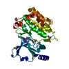
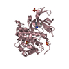
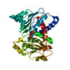
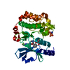
- PDB-4njd: Structure of p21-activated kinase 4 with a novel inhibitor KY-04031 -
+ Open data

- Basic information
| Entry | Database: PDB / ID: 4njd | ||||||
|---|---|---|---|---|---|---|---|
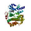
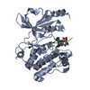
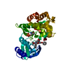
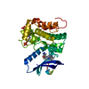
| Title | Structure of p21-activated kinase 4 with a novel inhibitor KY-04031 | ||||||
 Components Components | Serine/threonine-protein kinase PAK 4 | ||||||
 Keywords Keywords | Transferase/Transferase inhibitor /  Kinase / Transferase-Transferase inhibitor complex Kinase / Transferase-Transferase inhibitor complex | ||||||
| Function / homology |  Function and homology information Function and homology informationdendritic spine development / cadherin binding involved in cell-cell adhesion / Activation of RAC1 / RHOV GTPase cycle / RHOJ GTPase cycle / RHOQ GTPase cycle / regulation of MAPK cascade / RHOH GTPase cycle / CDC42 GTPase cycle / RHOU GTPase cycle ...dendritic spine development / cadherin binding involved in cell-cell adhesion / Activation of RAC1 / RHOV GTPase cycle / RHOJ GTPase cycle / RHOQ GTPase cycle / regulation of MAPK cascade / RHOH GTPase cycle / CDC42 GTPase cycle / RHOU GTPase cycle / RHOG GTPase cycle / cellular response to organic cyclic compound / RAC2 GTPase cycle / RAC3 GTPase cycle / negative regulation of endothelial cell apoptotic process / cytoskeleton organization / RAC1 GTPase cycle / regulation of cell growth / adherens junction / positive regulation of angiogenesis / cell migration / non-specific serine/threonine protein kinase / protein kinase activity / intracellular signal transduction / cell cycle / phosphorylation / focal adhesion / protein serine kinase activity / protein serine/threonine kinase activity / apoptotic process / Golgi apparatus / signal transduction / ATP binding / cytosol / cytoplasmSimilarity search - Function | ||||||
| Biological species |  Homo sapiens (human) Homo sapiens (human) | ||||||
| Method | X-RAY DIFFRACTION / SYNCHROTRON / MOLECULAR REPLACEMENT / Resolution: 2.5 Å | ||||||
 Authors Authors | Park, S. | ||||||
 Citation Citation | Journal: Cancer Lett. / Year: 2014 Title: Discovery and the structural basis of a novel p21-activated kinase 4 inhibitor. Authors: Ryu, B.J. / Kim, S. / Min, B. / Kim, K.Y. / Lee, J.S. / Park, W.J. / Lee, H. / Kim, S.H. / Park, S. | ||||||
| History |
|
- Structure visualization
| Structure viewer | Molecule: MolmilJmol/JSmol |
|---|
- Downloads & links
-Download
| PDBx/mmCIF format | 4njd.cif.gz | 128.8 KB | Display | PDBx/mmCIF format |
|---|---|---|---|---|
| PDB format | pdb4njd.ent.gz | 104.5 KB | Display | PDB format |
| PDBx/mmJSON format | 4njd.json.gz | Tree view | PDBx/mmJSON format | |
| Others |  Other downloads Other downloads |
-Validation report
| Arichive directory | https://data.pdbj.org/pub/pdb/validation_reports/nj/4njdftp://data.pdbj.org/pub/pdb/validation_reports/nj/4njd | HTTPS FTP |
|---|
-Related structure data
| Similar structure data |
|---|


-Links
 PDBj
PDBj


- Assembly
| Deposited unit | 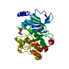
| ||||||||
|---|---|---|---|---|---|---|---|---|---|
| 1 |
| ||||||||
| Unit cell |
|
-Components
| #1: Protein | Mass: 33424.840 Da / Num. of mol.: 1 / Fragment: UNP residues 300-591 Source method: isolated from a genetically manipulated source Source: (gene. exp.) Homo sapiens (human) / Gene: PAK4 / Production host:  Escherichia coli (E. coli) Escherichia coli (E. coli)References: UniProt: O96013, non-specific serine/threonine protein kinase |
|---|---|
| #2: Chemical | ChemComp-NJD /   Mass: 400.437 Da / Num. of mol.: 1 / Source method: obtained synthetically / Formula: C21H20N8O Mass: 400.437 Da / Num. of mol.: 1 / Source method: obtained synthetically / Formula: C21H20N8O |
| #3: Water | ChemComp-HOH / Water Mass: 18.015 Da / Num. of mol.: 43 / Source method: isolated from a natural source / Formula: H2O Mass: 18.015 Da / Num. of mol.: 43 / Source method: isolated from a natural source / Formula: H2O |
-Experimental details
-Experiment
| Experiment | Method: X-RAY DIFFRACTION / Number of used crystals: 1 |
|---|
- Sample preparation
| Crystal | Density Matthews: 2.95 Å3/Da / Density % sol: 58.32 % |
|---|---|
| Crystal grow | Temperature: 297 K / Method: vapor diffusion, hanging drop / pH: 8.5 Details: 0.6M sodium potassium tartrate, 0.1M Tris, pH 8.5, VAPOR DIFFUSION, HANGING DROP, temperature 297K |
-Data collection
| Diffraction | Mean temperature: 100 K |
|---|---|
| Diffraction source | Source: SYNCHROTRON / Site: PAL/PLS  / Beamline: 5C (4A) / Wavelength: 1 Å / Beamline: 5C (4A) / Wavelength: 1 Å |
| Detector | Type: ADSC QUANTUM 270 / Detector: CCD / Date: Jun 22, 2013 |
| Radiation | Protocol: SINGLE WAVELENGTH / Monochromatic (M) / Laue (L): M / Scattering type: x-ray |
| Radiation wavelength | Wavelength: 1 Å / Relative weight: 1 |
| Reflection | Resolution: 2.5→50 Å / Num. obs: 13855 |
- Processing
| Software |
| ||||||||||||||||||||||||||||||||||||||||||||||||||||||||||||||||||||||||||||||||||||||||||||||||||||||||||||||||||||||||||||||||||||||||||||||||||||||||||||||||||||||||||||||||||||||
|---|---|---|---|---|---|---|---|---|---|---|---|---|---|---|---|---|---|---|---|---|---|---|---|---|---|---|---|---|---|---|---|---|---|---|---|---|---|---|---|---|---|---|---|---|---|---|---|---|---|---|---|---|---|---|---|---|---|---|---|---|---|---|---|---|---|---|---|---|---|---|---|---|---|---|---|---|---|---|---|---|---|---|---|---|---|---|---|---|---|---|---|---|---|---|---|---|---|---|---|---|---|---|---|---|---|---|---|---|---|---|---|---|---|---|---|---|---|---|---|---|---|---|---|---|---|---|---|---|---|---|---|---|---|---|---|---|---|---|---|---|---|---|---|---|---|---|---|---|---|---|---|---|---|---|---|---|---|---|---|---|---|---|---|---|---|---|---|---|---|---|---|---|---|---|---|---|---|---|---|---|---|---|---|
| Refinement | Method to determine structure: MOLECULAR REPLACEMENT / Resolution: 2.5→46.36 Å / Cor.coef. Fo:Fc: 0.923 / Cor.coef. Fo:Fc free: 0.868 / SU B: 24.461 / SU ML: 0.247 / Cross valid method: THROUGHOUT / ESU R: 0.498 / ESU R Free: 0.355 / Stereochemistry target values: MAXIMUM LIKELIHOOD / Details: HYDROGENS HAVE BEEN ADDED IN THE RIDING POSITIONS
| ||||||||||||||||||||||||||||||||||||||||||||||||||||||||||||||||||||||||||||||||||||||||||||||||||||||||||||||||||||||||||||||||||||||||||||||||||||||||||||||||||||||||||||||||||||||
| Solvent computation | Ion probe radii: 0.8 Å / Shrinkage radii: 0.8 Å / VDW probe radii: 1.4 Å / Solvent model: MASK | ||||||||||||||||||||||||||||||||||||||||||||||||||||||||||||||||||||||||||||||||||||||||||||||||||||||||||||||||||||||||||||||||||||||||||||||||||||||||||||||||||||||||||||||||||||||
| Displacement parameters | Biso mean: 67.198 Å2
| ||||||||||||||||||||||||||||||||||||||||||||||||||||||||||||||||||||||||||||||||||||||||||||||||||||||||||||||||||||||||||||||||||||||||||||||||||||||||||||||||||||||||||||||||||||||
| Refinement step | Cycle: LAST / Resolution: 2.5→46.36 Å
| ||||||||||||||||||||||||||||||||||||||||||||||||||||||||||||||||||||||||||||||||||||||||||||||||||||||||||||||||||||||||||||||||||||||||||||||||||||||||||||||||||||||||||||||||||||||
| Refine LS restraints |
|