 Movie
Movie Controller


+ Open data

- Basic information
| Entry | Database: PDB / ID: 4nby | ||||||
|---|---|---|---|---|---|---|---|
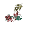
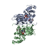
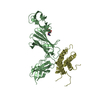
| Title | Crystal Structure of TcdA-A2 Bound to Two Molecules of A20.1 VHH | ||||||
 Components Components |
| ||||||
 Keywords Keywords |  IMMUNE SYSTEM / antibody-antigen complex IMMUNE SYSTEM / antibody-antigen complex | ||||||
| Function / homology | Cholin Binding / left handed beta-beta-3-solenoid / Ribbon / Immunoglobulins / Immunoglobulin-like / Sandwich / Mainly Beta / :  Function and homology information Function and homology information | ||||||
| Biological species |  Clostridium difficile (bacteria) Clostridium difficile (bacteria) Lama glama (llama) Lama glama (llama) | ||||||
| Method | X-RAY DIFFRACTION / SYNCHROTRON / MOLECULAR REPLACEMENT / Resolution: 2.08 Å | ||||||
 Authors Authors | Murase, T. / Eugenio, L. / Schorr, M. / Hussack, G. / Tanha, J. / Kitova, E. / Klassen, J.S. / Ng, K.K.S. | ||||||
 Citation Citation | Journal: J.Biol.Chem. / Year: 2014 Title: Structural Basis for Antibody Recognition in the Receptor-binding Domains of Toxins A and B from Clostridium difficile. Authors: Murase, T. / Eugenio, L. / Schorr, M. / Hussack, G. / Tanha, J. / Kitova, E.N. / Klassen, J.S. / Ng, K.K. | ||||||
| History |
|
- Structure visualization
| Structure viewer | Molecule: MolmilJmol/JSmol |
|---|
- Downloads & links
-Download
| PDBx/mmCIF format | 4nby.cif.gz | 118.3 KB | Display | PDBx/mmCIF format |
|---|---|---|---|---|
| PDB format | pdb4nby.ent.gz | 90 KB | Display | PDB format |
| PDBx/mmJSON format | 4nby.json.gz | Tree view | PDBx/mmJSON format | |
| Others |  Other downloads Other downloads |
-Validation report
| Arichive directory | https://data.pdbj.org/pub/pdb/validation_reports/nb/4nbyftp://data.pdbj.org/pub/pdb/validation_reports/nb/4nby | HTTPS FTP |
|---|
-Related structure data
| Related structure data |  4nbxC  4nbzC  4nc0C  4nc1C  4nc2C  2g7cS C: citing same article ( S: Starting model for refinement |
|---|---|
| Similar structure data |


-Links
 PDBj
PDBj

- Assembly
| Deposited unit | 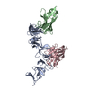
| ||||||||
|---|---|---|---|---|---|---|---|---|---|
| 1 |
| ||||||||
| Unit cell |
|
-Components
| #1: Protein | Mass: 29547.760 Da / Num. of mol.: 1 / Fragment: UNP residues 1-248 Source method: isolated from a genetically manipulated source Source: (gene. exp.) Clostridium difficile (bacteria) / Strain: 48489 / Gene: HMPREF0219_0762, TcdA / Production host: Escherichia coli (E. coli) / References: UniProt: D5RWT1 | ||
|---|---|---|---|
| #2: Antibody | Mass: 16828.449 Da / Num. of mol.: 2 Source method: isolated from a genetically manipulated source Source: (gene. exp.) Lama glama (llama) / Gene: VHH / Production host: Escherichia coli (E. coli)#3: Water | ChemComp-HOH / | Water Mass: 18.015 Da / Num. of mol.: 408 / Source method: isolated from a natural source / Formula: H2O Mass: 18.015 Da / Num. of mol.: 408 / Source method: isolated from a natural source / Formula: H2O |
-Experimental details
-Experiment
| Experiment | Method: X-RAY DIFFRACTION / Number of used crystals: 1 |
|---|
- Sample preparation
| Crystal | Density Matthews: 3.03 Å3/Da / Density % sol: 59.42 % |
|---|---|
| Crystal grow | Temperature: 295 K / Method: vapor diffusion, hanging drop / pH: 7.5 Details: 1.1 M Ammonium sulfate, 0.1 M Tris-Cl, pH 7.5, 0.1 M TMAO, VAPOR DIFFUSION, HANGING DROP, temperature 295K |
-Data collection
| Diffraction | Mean temperature: 100 K |
|---|---|
| Diffraction source | Source: SYNCHROTRON / Site: CLSI  / Beamline: 08B1-1 / Wavelength: 0.98 Å / Beamline: 08B1-1 / Wavelength: 0.98 Å |
| Detector | Type: MARMOSAIC 300 mm CCD / Detector: CCD / Date: Mar 16, 2012 / Details: mirrors |
| Radiation | Monochromator: Si(111) double crystal monochromator / Protocol: SINGLE WAVELENGTH / Monochromatic (M) / Laue (L): M / Scattering type: x-ray |
| Radiation wavelength | Wavelength: 0.98 Å / Relative weight: 1 |
| Reflection | Resolution: 2.08→40 Å / Num. all: 48737 / Num. obs: 48737 / % possible obs: 99.9 % / Observed criterion σ(F): -3 / Observed criterion σ(I): -3 / Redundancy: 14.1 % / Biso Wilson estimate: 36.9 Å2 / Rmerge(I) obs: 0.085 / Rsym value: 0.085 / Net I/σ(I): 23.4 |
| Reflection shell | Resolution: 2.08→2.13 Å / Redundancy: 14.5 % / Rmerge(I) obs: 0.692 / Mean I/σ(I) obs: 5.2 / Num. unique all: 3513 / Rsym value: 0.692 / % possible all: 99.9 |
- Processing
| Software |
| |||||||||||||||||||||||||||||||||||||||||||||
|---|---|---|---|---|---|---|---|---|---|---|---|---|---|---|---|---|---|---|---|---|---|---|---|---|---|---|---|---|---|---|---|---|---|---|---|---|---|---|---|---|---|---|---|---|---|---|
| Refinement | Method to determine structure: MOLECULAR REPLACEMENT Starting model: PDB entry 2G7C Resolution: 2.08→40 Å / Cor.coef. Fo:Fc: 0.942 / Cor.coef. Fo:Fc free: 0.915 / SU B: 4.121 / SU ML: 0.112 / Cross valid method: THROUGHOUT / σ(F): 0 / σ(I): 0 / ESU R: 0.17 / ESU R Free: 0.164 / Stereochemistry target values: MAXIMUM LIKELIHOOD
| |||||||||||||||||||||||||||||||||||||||||||||
| Solvent computation | Ion probe radii: 0.8 Å / Shrinkage radii: 0.8 Å / VDW probe radii: 1.4 Å / Solvent model: BABINET MODEL WITH MASK | |||||||||||||||||||||||||||||||||||||||||||||
| Displacement parameters | Biso mean: 34.296 Å2
| |||||||||||||||||||||||||||||||||||||||||||||
| Refinement step | Cycle: LAST / Resolution: 2.08→40 Å
| |||||||||||||||||||||||||||||||||||||||||||||
| Refine LS restraints |
| |||||||||||||||||||||||||||||||||||||||||||||
| LS refinement shell | Resolution: 2.08→2.134 Å / Total num. of bins used: 20
|
