 Movie
Movie Controller

[English] 日本語
 Yorodumi
- PDB-4nar: Crystal Structure of the Q9WYS3 protein from Thermotoga maritima.... -
+ Open data

- Basic information
| Entry | Database: PDB / ID: 4nar | ||||||
|---|---|---|---|---|---|---|---|
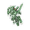
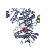
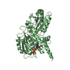
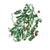
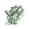
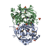
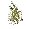
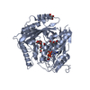
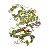
| Title | Crystal Structure of the Q9WYS3 protein from Thermotoga maritima. Northeast Structural Genomics Consortium Target VR152 | ||||||
 Components Components | Putative uronate isomerase | ||||||
 Keywords Keywords |  ISOMERASE / Structural Genomics / PSI-Biology / Protein Structure Initiative / Northeast Structural Genomics Consortium / NESG / VR152 / PF09861 / DUF2088 ISOMERASE / Structural Genomics / PSI-Biology / Protein Structure Initiative / Northeast Structural Genomics Consortium / NESG / VR152 / PF09861 / DUF2088 | ||||||
| Function / homology |  Function and homology information Function and homology information | ||||||
| Biological species |   Thermotoga maritima (bacteria) Thermotoga maritima (bacteria) | ||||||
| Method | X-RAY DIFFRACTION / SYNCHROTRON / SAD / Resolution: 2.388 Å | ||||||
 Authors Authors | Vorobiev, S. / Lew, S. / Seetharaman, J. / Chi, Y. / Xiao, R. / Maglaqui, M. / Lee, D. / Everett, J.K. / Acton, T.B. / Montelione, G.T. ...Vorobiev, S. / Lew, S. / Seetharaman, J. / Chi, Y. / Xiao, R. / Maglaqui, M. / Lee, D. / Everett, J.K. / Acton, T.B. / Montelione, G.T. / Tong, L. / Hunt, J.F. / Northeast Structural Genomics Consortium (NESG) | ||||||
 Citation Citation | Journal: To be Published Title: Crystal Structure of the Q9WYS3 protein from Thermotoga maritima. Authors: Vorobiev, S. / Lew, S. / Seetharaman, J. / Chi, Y. / Xiao, R. / Maglaqui, M. / Lee, D. / Everett, J.K. / Acton, T.B. / Montelione, G.T. / Tong, L. / Hunt, J.F. | ||||||
| History |
|
- Structure visualization
| Structure viewer | Molecule: MolmilJmol/JSmol |
|---|
- Downloads & links
-Download
| PDBx/mmCIF format | 4nar.cif.gz | 180.5 KB | Display | PDBx/mmCIF format |
|---|---|---|---|---|
| PDB format | pdb4nar.ent.gz | 150.4 KB | Display | PDB format |
| PDBx/mmJSON format | 4nar.json.gz | Tree view | PDBx/mmJSON format | |
| Others |  Other downloads Other downloads |
-Validation report
| Arichive directory | https://data.pdbj.org/pub/pdb/validation_reports/na/4narftp://data.pdbj.org/pub/pdb/validation_reports/na/4nar | HTTPS FTP |
|---|
-Related structure data
| Similar structure data | |
|---|---|
| Other databases |

-Links
 PDBj
PDBj- Assembly
| Deposited unit | 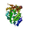
| ||||||||
|---|---|---|---|---|---|---|---|---|---|
| 1 |
| ||||||||
| 2 | 
| ||||||||
| Unit cell |
| ||||||||
| Components on special symmetry positions |
|
-Components
| #1: Protein | Mass: 50666.281 Da / Num. of mol.: 1 Source method: isolated from a genetically manipulated source Source: (gene. exp.) Thermotoga maritima (bacteria) / Strain: ATCC 43589/MSB8/DSM 3109/JCM 10099 / Gene: Tmari_0439, TM_0442 / Production host: Escherichia coli (E. coli) / Strain (production host): BL21(DE3)+Magic / References: UniProt: Q9WYS3 | ||||
|---|---|---|---|---|---|
| #2: Chemical | Sulfate  Mass: 96.063 Da / Num. of mol.: 3 / Source method: obtained synthetically / Formula: SO4 Mass: 96.063 Da / Num. of mol.: 3 / Source method: obtained synthetically / Formula: SO4#3: Chemical | ChemComp-ACT / | Acetate  Mass: 59.044 Da / Num. of mol.: 1 / Source method: obtained synthetically / Formula: C2H3O2 Mass: 59.044 Da / Num. of mol.: 1 / Source method: obtained synthetically / Formula: C2H3O2#4: Water | ChemComp-HOH / | Water Mass: 18.015 Da / Num. of mol.: 160 / Source method: isolated from a natural source / Formula: H2O Mass: 18.015 Da / Num. of mol.: 160 / Source method: isolated from a natural source / Formula: H2O |
-Experimental details
-Experiment
| Experiment | Method: X-RAY DIFFRACTION / Number of used crystals: 1 |
|---|
- Sample preparation
| Crystal | Density Matthews: 2.19 Å3/Da / Density % sol: 43.87 % |
|---|---|
| Crystal grow | Temperature: 291 K / Method: microbatch crystallization under oil / pH: 4.6 Details: 30% PEG MME 2000, 0.2 M ammonium sulfate, 0.1 M sodium acetate, pH 4.6, Microbatch crystallization under oil, temperature 291K |
-Data collection
| Diffraction | Mean temperature: 100 K |
|---|---|
| Diffraction source | Source: SYNCHROTRON / Site: NSLS  / Beamline: X4C / Wavelength: 0.97915 Å / Beamline: X4C / Wavelength: 0.97915 Å |
| Detector | Type: MAR CCD 165 mm / Detector: CCD / Date: Oct 10, 2013 |
| Radiation | Monochromator: Si 111 CHANNEL / Protocol: SINGLE WAVELENGTH / Monochromatic (M) / Laue (L): M / Scattering type: x-ray |
| Radiation wavelength | Wavelength: 0.97915 Å / Relative weight: 1 |
| Reflection | Resolution: 2.39→50 Å / Num. all: 18378 / Num. obs: 18360 / % possible obs: 99.9 % / Observed criterion σ(F): 0 / Observed criterion σ(I): 0 / Redundancy: 9.9 % / Biso Wilson estimate: 26.84 Å2 / Rmerge(I) obs: 0.122 / Net I/σ(I): 22.8 |
| Reflection shell | Resolution: 2.39→2.48 Å / Redundancy: 8 % / Rmerge(I) obs: 0.338 / Mean I/σ(I) obs: 4.7 / Num. unique all: 1807 / % possible all: 98.9 |
- Processing
| Software |
| |||||||||||||||||||||||||||||||||||||||||||||||||||||||||||||||||||||||||||||||||||||||||||
|---|---|---|---|---|---|---|---|---|---|---|---|---|---|---|---|---|---|---|---|---|---|---|---|---|---|---|---|---|---|---|---|---|---|---|---|---|---|---|---|---|---|---|---|---|---|---|---|---|---|---|---|---|---|---|---|---|---|---|---|---|---|---|---|---|---|---|---|---|---|---|---|---|---|---|---|---|---|---|---|---|---|---|---|---|---|---|---|---|---|---|---|---|
| Refinement | Method to determine structure: SAD / Resolution: 2.388→47.023 Å / Occupancy max: 1 / Occupancy min: 0.21 / SU ML: 0.62 / Cross valid method: THROUGHOUT / σ(F): 1.35 / Phase error: 21.89 / Stereochemistry target values: ML
| |||||||||||||||||||||||||||||||||||||||||||||||||||||||||||||||||||||||||||||||||||||||||||
| Solvent computation | Shrinkage radii: 0.9 Å / VDW probe radii: 1.11 Å / Solvent model: FLAT BULK SOLVENT MODEL / Bsol: 38.168 Å2 / ksol: 0.382 e/Å3 | |||||||||||||||||||||||||||||||||||||||||||||||||||||||||||||||||||||||||||||||||||||||||||
| Displacement parameters | Biso max: 166.92 Å2 / Biso mean: 31.386 Å2 / Biso min: 10.78 Å2
| |||||||||||||||||||||||||||||||||||||||||||||||||||||||||||||||||||||||||||||||||||||||||||
| Refinement step | Cycle: LAST / Resolution: 2.388→47.023 Å
| |||||||||||||||||||||||||||||||||||||||||||||||||||||||||||||||||||||||||||||||||||||||||||
| Refine LS restraints |
| |||||||||||||||||||||||||||||||||||||||||||||||||||||||||||||||||||||||||||||||||||||||||||
| LS refinement shell | Refine-ID: X-RAY DIFFRACTION / Total num. of bins used: 12
| |||||||||||||||||||||||||||||||||||||||||||||||||||||||||||||||||||||||||||||||||||||||||||
| Refinement TLS params. | Method: refined / Origin x: -17.1145 Å / Origin y: 23.9907 Å / Origin z: -13.4147 Å
| |||||||||||||||||||||||||||||||||||||||||||||||||||||||||||||||||||||||||||||||||||||||||||
| Refinement TLS group | Selection details: chain A |
