 Movie
Movie Controller


+ Open data

- Basic information
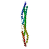
| Entry | Database: PDB / ID: 4n3y | ||||||
|---|---|---|---|---|---|---|---|
| Title | Crystal structure of Rabex-5CC and Rabaptin-5C21 complex | ||||||
 Components Components |
| ||||||
 Keywords Keywords |  ENDOCYTOSIS / Rab5 / Rabex-5 / Rabaptin-5 / GEF activity / early endosome ENDOCYTOSIS / Rab5 / Rabex-5 / Rabaptin-5 / GEF activity / early endosome | ||||||
| Function / homology |  Function and homology information Function and homology informationdendritic transport / negative regulation of Kit signaling pathway / mast cell migration / regulation of Fc receptor mediated stimulatory signaling pathway / protein localization to ciliary membrane / Kit signaling pathway / negative regulation of mast cell degranulation / negative regulation of mast cell activation / negative regulation of leukocyte migration / negative regulation of receptor-mediated endocytosis ...dendritic transport / negative regulation of Kit signaling pathway / mast cell migration / regulation of Fc receptor mediated stimulatory signaling pathway / protein localization to ciliary membrane / Kit signaling pathway / negative regulation of mast cell degranulation / negative regulation of mast cell activation / negative regulation of leukocyte migration / negative regulation of receptor-mediated endocytosis / negative regulation of mast cell cytokine production / Golgi to plasma membrane transport / RAB GEFs exchange GTP for GDP on RABs / negative regulation of Ras protein signal transduction / protein targeting to membrane / TBC/RABGAPs / mast cell degranulation / negative regulation of interleukin-6 production / endocytic vesicle / vesicle-mediated transport / receptor-mediated endocytosis / GTPase activator activity / guanyl-nucleotide exchange factor activity / negative regulation of protein phosphorylation / growth factor activity / recycling endosome / small GTPase binding / negative regulation of inflammatory response / endocytosis / ubiquitin protein ligase activity / protein transport / early endosome membrane / Ras protein signal transduction / membrane fusion / early endosome / endosome / protein domain specific binding / intracellular membrane-bounded organelle / apoptotic process / dendrite / nucleolus / protein homodimerization activity / protein-containing complex / DNA binding / zinc ion binding / cytosolSimilarity search - Function | ||||||
| Biological species |  Homo sapiens (human) Homo sapiens (human) | ||||||
| Method | X-RAY DIFFRACTION / SYNCHROTRON / MOLECULAR REPLACEMENT / molecular replacement / Resolution: 2.2 Å | ||||||
 Authors Authors | Zhang, Z. / Zhang, T. / Ding, J. | ||||||
 Citation Citation | Journal: Elife / Year: 2014 Title: Molecular mechanism for Rabex-5 GEF activation by Rabaptin-5 Authors: Zhang, Z. / Zhang, T. / Wang, S. / Gong, Z. / Tang, C. / Chen, J. / Ding, J. | ||||||
| History |
|
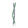
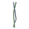
- Structure visualization
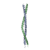
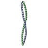
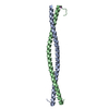
| Structure viewer | Molecule: MolmilJmol/JSmol |
|---|
- Downloads & links
-Download
| PDBx/mmCIF format | 4n3y.cif.gz | 104.4 KB | Display | PDBx/mmCIF format |
|---|---|---|---|---|
| PDB format | pdb4n3y.ent.gz | 81.7 KB | Display | PDB format |
| PDBx/mmJSON format | 4n3y.json.gz | Tree view | PDBx/mmJSON format | |
| Others |  Other downloads Other downloads |
-Validation report
| Arichive directory | https://data.pdbj.org/pub/pdb/validation_reports/n3/4n3yftp://data.pdbj.org/pub/pdb/validation_reports/n3/4n3y | HTTPS FTP |
|---|
-Related structure data
| Related structure data |  4n3xC  4n3zC 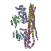 4q9uC  1x79S C: citing same article ( S: Starting model for refinement |
|---|---|
| Similar structure data |


-Links
 PDBj
PDBj

- Assembly
| Deposited unit | 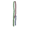
| ||||||||
|---|---|---|---|---|---|---|---|---|---|
| 1 |
| ||||||||
| Unit cell |
|
-Components
| #1: Protein/peptide | Mass: 5261.896 Da / Num. of mol.: 1 / Fragment: UNP residues 630-672 Source method: isolated from a genetically manipulated source Source: (gene. exp.) Homo sapiens (human) / Gene: RABEX5 / Plasmid: pETDuet1 / Production host:  Escherichia coli (E. coli) / Strain (production host): BL21(DE3) codon plus / References: UniProt: Q9UJ41 Escherichia coli (E. coli) / Strain (production host): BL21(DE3) codon plus / References: UniProt: Q9UJ41 | ||
|---|---|---|---|
| #2: Protein | Mass: 10701.068 Da / Num. of mol.: 2 / Fragment: UNP residues 552-642 Source method: isolated from a genetically manipulated source Source: (gene. exp.) Homo sapiens (human) / Gene: RABEP1 / Plasmid: pETDuet1 / Production host: Escherichia coli (E. coli) / Strain (production host): BL21(DE3) codon plus / References: UniProt: Q15276#3: Water | ChemComp-HOH / | Water Mass: 18.015 Da / Num. of mol.: 187 / Source method: isolated from a natural source / Formula: H2O Mass: 18.015 Da / Num. of mol.: 187 / Source method: isolated from a natural source / Formula: H2O |
-Experimental details
-Experiment
| Experiment | Method: X-RAY DIFFRACTION / Number of used crystals: 1 |
|---|
- Sample preparation
| Crystal | Density Matthews: 2.57 Å3/Da / Density % sol: 52.13 % |
|---|---|
| Crystal grow | Temperature: 289 K / Method: vapor diffusion, hanging drop Details: 0.15M MgAc2, 20% PEG3350, VAPOR DIFFUSION, HANGING DROP, temperature 289K |
-Data collection
| Diffraction | Mean temperature: 100 K |
|---|---|
| Diffraction source | Source: SYNCHROTRON / Site: SSRF  / Beamline: BL17U / Wavelength: 1 Å / Beamline: BL17U / Wavelength: 1 Å |
| Detector | Type: ADSC QUANTUM 315r / Detector: CCD / Date: May 21, 2012 |
| Radiation | Protocol: SINGLE WAVELENGTH / Monochromatic (M) / Laue (L): M / Scattering type: x-ray |
| Radiation wavelength | Wavelength: 1 Å / Relative weight: 1 |
| Reflection | Resolution: 2.2→50 Å / Num. obs: 13816 / % possible obs: 97.7 % / Redundancy: 3.4 % / Rmerge(I) obs: 0.06 / Net I/σ(I): 21.2 |
| Reflection shell | Resolution: 2.2→2.28 Å / Redundancy: 3 % / Rmerge(I) obs: 0.28 / Mean I/σ(I) obs: 3.4 / Num. unique all: 13816 / % possible all: 85.8 |
-Phasing
| Phasing | Method: molecular replacement |
|---|
- Processing
| Software |
| ||||||||||||||||||||||||||||||||||||||||||||||||||||||||||||||||||||||||||||||||||||||||||||||||||||
|---|---|---|---|---|---|---|---|---|---|---|---|---|---|---|---|---|---|---|---|---|---|---|---|---|---|---|---|---|---|---|---|---|---|---|---|---|---|---|---|---|---|---|---|---|---|---|---|---|---|---|---|---|---|---|---|---|---|---|---|---|---|---|---|---|---|---|---|---|---|---|---|---|---|---|---|---|---|---|---|---|---|---|---|---|---|---|---|---|---|---|---|---|---|---|---|---|---|---|---|---|---|
| Refinement | Method to determine structure: MOLECULAR REPLACEMENT Starting model: 1X79 Resolution: 2.2→15.03 Å / Cor.coef. Fo:Fc: 0.961 / Cor.coef. Fo:Fc free: 0.948 / Occupancy max: 1 / Occupancy min: 1 / SU B: 17.797 / SU ML: 0.187 / Cross valid method: THROUGHOUT / σ(F): 0 / ESU R Free: 0.197 / Stereochemistry target values: MAXIMUM LIKELIHOOD Details: HYDROGENS HAVE BEEN ADDED IN THE RIDING POSITIONS U VALUES: RESIDUAL ONLY
| ||||||||||||||||||||||||||||||||||||||||||||||||||||||||||||||||||||||||||||||||||||||||||||||||||||
| Solvent computation | Ion probe radii: 0.8 Å / Shrinkage radii: 0.8 Å / VDW probe radii: 1.2 Å / Solvent model: MASK | ||||||||||||||||||||||||||||||||||||||||||||||||||||||||||||||||||||||||||||||||||||||||||||||||||||
| Displacement parameters | Biso max: 162.73 Å2 / Biso mean: 58.5336 Å2 / Biso min: 22.48 Å2
| ||||||||||||||||||||||||||||||||||||||||||||||||||||||||||||||||||||||||||||||||||||||||||||||||||||
| Refinement step | Cycle: LAST / Resolution: 2.2→15.03 Å
| ||||||||||||||||||||||||||||||||||||||||||||||||||||||||||||||||||||||||||||||||||||||||||||||||||||
| Refine LS restraints |
| ||||||||||||||||||||||||||||||||||||||||||||||||||||||||||||||||||||||||||||||||||||||||||||||||||||
| LS refinement shell | Resolution: 2.203→2.26 Å / Total num. of bins used: 20
| ||||||||||||||||||||||||||||||||||||||||||||||||||||||||||||||||||||||||||||||||||||||||||||||||||||
| Refinement TLS params. | Method: refined / Refine-ID: X-RAY DIFFRACTION
| ||||||||||||||||||||||||||||||||||||||||||||||||||||||||||||||||||||||||||||||||||||||||||||||||||||
| Refinement TLS group |
|
