| Entry | Database: PDB / ID: 4n1x
|
|---|
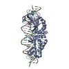
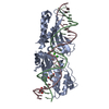
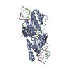
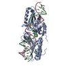
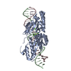
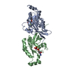
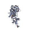
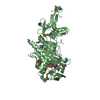
| Title | Structure of a putative peptidoglycan glycosyltransferase from Atopobium parvulum in complex with penicillin G |
|---|
 Components Components | Peptidoglycan glycosyltransferase |
|---|
 Keywords Keywords | Transferase/Antibiotic / Structural Genomics / PSI-Biology / Midwest Center for Structural Genomics / MCSG / Susceptibility to Known Mtb Inhibitors / MTBI / glycosyltransferase / penicillin G / Transferase-Antibiotic Complex / Structures of Mtb Proteins Conferring Susceptibility to Known Mtb Inhibitors |
|---|
| Function / homology |  Function and homology information Function and homology information |
|---|
| Biological species |  Atopobium parvulum (bacteria) Atopobium parvulum (bacteria) |
|---|
| Method | X-RAY DIFFRACTION / SYNCHROTRON / MOLECULAR REPLACEMENT / Resolution: 2 Å |
|---|
 Authors Authors | Filippova, E.V. / Minasov, G. / Shuvalova, L. / Kiryukhina, O. / Babnigg, G. / Rubin, E. / Sacchettini, J. / Joachimiak, A. / Anderson, W.F. / Midwest Center for Structural Genomics (MCSG) / Structures of Mtb Proteins Conferring Susceptibility to Known Mtb Inhibitors (MTBI) |
|---|
 Citation Citation | Journal: To be Published
Title: Structure of a putative peptidoglycan glycosyltransferase from Atopobium parvulum in complex with penicillin G
Authors: Filippova, E.V. / Minasov, G. / Shuvalova, L. / Kiryukhina, O. / Babnigg, G. / Rubin, E. / Sacchettini, J. / Joachimiak, A. / Anderson, W.F. |
|---|
| History | | Deposition | Oct 4, 2013 | Deposition site: RCSB / Processing site: RCSB |
|---|
| Revision 1.0 | Oct 30, 2013 | Provider: repository / Type: Initial release |
|---|
| Revision 1.1 | Aug 13, 2014 | Group: Structure summary |
|---|
| Revision 1.2 | Nov 15, 2017 | Group: Refinement description / Category: software / Item: _software.name |
|---|
| Revision 1.3 | Sep 20, 2023 | Group: Data collection / Database references ...Data collection / Database references / Derived calculations / Refinement description
Category: chem_comp_atom / chem_comp_bond ...chem_comp_atom / chem_comp_bond / database_2 / pdbx_initial_refinement_model / struct_conn / struct_ref_seq_dif / struct_site
Item: _database_2.pdbx_DOI / _database_2.pdbx_database_accession ..._database_2.pdbx_DOI / _database_2.pdbx_database_accession / _struct_conn.pdbx_dist_value / _struct_conn.pdbx_leaving_atom_flag / _struct_conn.pdbx_ptnr2_label_alt_id / _struct_conn.ptnr1_auth_asym_id / _struct_conn.ptnr1_auth_comp_id / _struct_conn.ptnr1_auth_seq_id / _struct_conn.ptnr1_label_asym_id / _struct_conn.ptnr1_label_atom_id / _struct_conn.ptnr1_label_comp_id / _struct_conn.ptnr1_label_seq_id / _struct_conn.ptnr2_auth_asym_id / _struct_conn.ptnr2_auth_comp_id / _struct_conn.ptnr2_auth_seq_id / _struct_conn.ptnr2_label_asym_id / _struct_conn.ptnr2_label_atom_id / _struct_conn.ptnr2_label_comp_id / _struct_conn.ptnr2_label_seq_id / _struct_ref_seq_dif.details / _struct_site.pdbx_auth_asym_id / _struct_site.pdbx_auth_comp_id / _struct_site.pdbx_auth_seq_id |
|---|
| Revision 1.4 | Dec 6, 2023 | Group: Data collection / Category: chem_comp_atom / chem_comp_bond / Item: _chem_comp_atom.atom_id / _chem_comp_bond.atom_id_2 |
|---|
|
|---|
 Movie
Movie Controller

 Yorodumi
 Open data
 Basic information
 Structure visualization
 Downloads & links
 Other downloads
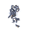


 PDBj
PDBj Assembly
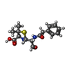
 Mass: 336.406 Da / Num. of mol.: 2 / Source method: obtained synthetically / Formula: C16H20N2O4S / Comment: antibiotic*YM
 Mass: 18.015 Da / Num. of mol.: 800 / Source method: isolated from a natural source / Formula: H2O
 Sample preparation
 / Beamline: 21-ID-F / Wavelength: 0.97872 Å
 Processing