 Movie
Movie Controller

[English] 日本語
 Yorodumi
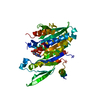
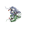
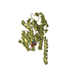
- PDB-4mym: Crystal structure of a glyoxalase/ bleomycin resistance protein/ ... -
+ Open data

- Basic information
| Entry | Database: PDB / ID: 4mym | ||||||
|---|---|---|---|---|---|---|---|
| Title | Crystal structure of a glyoxalase/ bleomycin resistance protein/ dioxygenase from Nocardioides | ||||||
 Components Components | Glyoxalase/bleomycin resistance protein/dioxygenase | ||||||
 Keywords Keywords |  OXIDOREDUCTASE / PSI-Biology / NYSGRC / Structural Genomics / New York Structural Genomics Research Consortium / metalloproteins / antibiotic resistance proteins OXIDOREDUCTASE / PSI-Biology / NYSGRC / Structural Genomics / New York Structural Genomics Research Consortium / metalloproteins / antibiotic resistance proteins | ||||||
| Function / homology |  Function and homology information Function and homology information | ||||||
| Biological species |  Nocardioides sp. (bacteria) Nocardioides sp. (bacteria) | ||||||
| Method | X-RAY DIFFRACTION / SYNCHROTRON / SAD / Resolution: 1.9 Å | ||||||
 Authors Authors | Eswaramoorthy, S. / Chamala, S. / Evans, B. / Foti, F. / Gizzi, A. / Hillerich, B. / Kar, A. / Lafleur, J. / Seidel, R. / Villigas, G. ...Eswaramoorthy, S. / Chamala, S. / Evans, B. / Foti, F. / Gizzi, A. / Hillerich, B. / Kar, A. / Lafleur, J. / Seidel, R. / Villigas, G. / Zencheck, W. / AL Obaidi, N. / Almo, S.C. / Swaminathan, S. / New York Structural Genomics Research Consortium (NYSGRC) | ||||||
 Citation Citation | Journal: To be Published Title: Crystal structure of a glyoxalase/ bleomycin resistance protein/ dioxygenase from Nocardioides. Authors: Eswaramoorthy, S. / Almo, S.C. / Swaminathan, S. | ||||||
| History |
|
- Structure visualization
| Structure viewer | Molecule: MolmilJmol/JSmol |
|---|
- Downloads & links
-Download
| PDBx/mmCIF format | 4mym.cif.gz | 41.4 KB | Display | PDBx/mmCIF format |
|---|---|---|---|---|
| PDB format | pdb4mym.ent.gz | 31.3 KB | Display | PDB format |
| PDBx/mmJSON format | 4mym.json.gz | Tree view | PDBx/mmJSON format | |
| Others |  Other downloads Other downloads |
-Validation report
| Arichive directory | https://data.pdbj.org/pub/pdb/validation_reports/my/4mymftp://data.pdbj.org/pub/pdb/validation_reports/my/4mym | HTTPS FTP |
|---|
-Related structure data
| Similar structure data | |
|---|---|
| Other databases |


-Links
 PDBj
PDBj- Assembly
| Deposited unit | 
| ||||||||
|---|---|---|---|---|---|---|---|---|---|
| 1 | 
| ||||||||
| Unit cell |
|
-Components
| #1: Protein | Mass: 19770.479 Da / Num. of mol.: 1 Source method: isolated from a genetically manipulated source Source: (gene. exp.) Nocardioides sp. (bacteria) / Strain: BAA-499 / JS614 / Gene: Noca_4190 / Plasmid: pET3A / Production host: Escherichia coli (E. coli) / Strain (production host): BL21 / References: UniProt: A1SPF2 |
|---|---|
| #2: Chemical | ChemComp-BTB / Bis-tris methane  Mass: 209.240 Da / Num. of mol.: 1 / Source method: obtained synthetically / Formula: C8H19NO5 / Comment: pH buffer*YM Mass: 209.240 Da / Num. of mol.: 1 / Source method: obtained synthetically / Formula: C8H19NO5 / Comment: pH buffer*YM |
| #3: Water | ChemComp-HOH / Water Mass: 18.015 Da / Num. of mol.: 52 / Source method: isolated from a natural source / Formula: H2O Mass: 18.015 Da / Num. of mol.: 52 / Source method: isolated from a natural source / Formula: H2O |
-Experimental details
-Experiment
| Experiment | Method: X-RAY DIFFRACTION / Number of used crystals: 1 |
|---|
- Sample preparation
| Crystal | Density Matthews: 2.31 Å3/Da / Density % sol: 46.73 % |
|---|---|
| Crystal grow | Temperature: 293 K / Method: vapor diffusion, sitting drop / pH: 5.5 Details: 25% PEG3350, 0.1M Bis-Tris, 0.2M NaCl, pH 5.5, VAPOR DIFFUSION, SITTING DROP, temperature 293K |
-Data collection
| Diffraction | Mean temperature: 100 K |
|---|---|
| Diffraction source | Source: SYNCHROTRON / Site: NSLS  / Beamline: X29A / Wavelength: 0.9792 Å / Beamline: X29A / Wavelength: 0.9792 Å |
| Detector | Type: ADSC QUANTUM 315 / Detector: CCD / Date: Sep 25, 2013 |
| Radiation | Monochromator: Si 111 CHANNEL / Protocol: SINGLE WAVELENGTH / Monochromatic (M) / Laue (L): M / Scattering type: x-ray |
| Radiation wavelength | Wavelength: 0.9792 Å / Relative weight: 1 |
| Reflection | Resolution: 1.9→50 Å / Num. all: 14825 / Num. obs: 14825 / % possible obs: 99.9 % / Observed criterion σ(F): 0 / Observed criterion σ(I): 0 / Redundancy: 13.8 % / Biso Wilson estimate: 20.012 Å2 / Rmerge(I) obs: 0.092 / Net I/σ(I): 11 |
| Reflection shell | Resolution: 1.9→1.96 Å / Redundancy: 12.6 % / Rmerge(I) obs: 0.455 / Num. unique all: 1228 / % possible all: 99.9 |
- Processing
| Software |
| ||||||||||||||||||||||||||||||||||||||||||||||||||||||||||||||||||||||||||||||||||||||||||||||||||||||||||||||||||||||||||||||||||||||||||||||||||||||||||||||||||||||||||||||||||||||
|---|---|---|---|---|---|---|---|---|---|---|---|---|---|---|---|---|---|---|---|---|---|---|---|---|---|---|---|---|---|---|---|---|---|---|---|---|---|---|---|---|---|---|---|---|---|---|---|---|---|---|---|---|---|---|---|---|---|---|---|---|---|---|---|---|---|---|---|---|---|---|---|---|---|---|---|---|---|---|---|---|---|---|---|---|---|---|---|---|---|---|---|---|---|---|---|---|---|---|---|---|---|---|---|---|---|---|---|---|---|---|---|---|---|---|---|---|---|---|---|---|---|---|---|---|---|---|---|---|---|---|---|---|---|---|---|---|---|---|---|---|---|---|---|---|---|---|---|---|---|---|---|---|---|---|---|---|---|---|---|---|---|---|---|---|---|---|---|---|---|---|---|---|---|---|---|---|---|---|---|---|---|---|---|
| Refinement | Method to determine structure: SAD / Resolution: 1.9→38.03 Å / Cor.coef. Fo:Fc: 0.957 / Cor.coef. Fo:Fc free: 0.939 / SU B: 3.207 / SU ML: 0.093 / Cross valid method: THROUGHOUT / ESU R: 0.134 / ESU R Free: 0.126 / Stereochemistry target values: MAXIMUM LIKELIHOOD / Details: HYDROGENS HAVE BEEN ADDED IN THE RIDING POSITIONS
| ||||||||||||||||||||||||||||||||||||||||||||||||||||||||||||||||||||||||||||||||||||||||||||||||||||||||||||||||||||||||||||||||||||||||||||||||||||||||||||||||||||||||||||||||||||||
| Solvent computation | Ion probe radii: 0.8 Å / Shrinkage radii: 0.8 Å / VDW probe radii: 1.2 Å / Solvent model: MASK | ||||||||||||||||||||||||||||||||||||||||||||||||||||||||||||||||||||||||||||||||||||||||||||||||||||||||||||||||||||||||||||||||||||||||||||||||||||||||||||||||||||||||||||||||||||||
| Displacement parameters | Biso mean: 25.849 Å2
| ||||||||||||||||||||||||||||||||||||||||||||||||||||||||||||||||||||||||||||||||||||||||||||||||||||||||||||||||||||||||||||||||||||||||||||||||||||||||||||||||||||||||||||||||||||||
| Refinement step | Cycle: LAST / Resolution: 1.9→38.03 Å
| ||||||||||||||||||||||||||||||||||||||||||||||||||||||||||||||||||||||||||||||||||||||||||||||||||||||||||||||||||||||||||||||||||||||||||||||||||||||||||||||||||||||||||||||||||||||
| Refine LS restraints |
|