 Movie
Movie Controller

[English] 日本語
 Yorodumi
- PDB-4mxt: Crystal structure of an Outer-membrane lipoprotein carrier protei... -
+ Open data

- Basic information
| Entry | Database: PDB / ID: 4mxt | ||||||
|---|---|---|---|---|---|---|---|
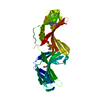
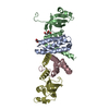
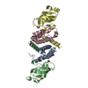
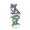
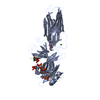
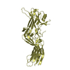
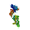
| Title | Crystal structure of an Outer-membrane lipoprotein carrier protein (BACUNI_04723) from Bacteroides uniformis ATCC 8492 at 1.40 A resolution | ||||||
 Components Components | Uncharacterized protein | ||||||
 Keywords Keywords |  PROTEIN TRANSPORT / LolA-like prokaryotic lipoproteins and lipoprotein localization factors fold / Structural Genomics / Joint Center for Structural Genomics / JCSG / Protein Structure Initiative / PSI-BIOLOGY PROTEIN TRANSPORT / LolA-like prokaryotic lipoproteins and lipoprotein localization factors fold / Structural Genomics / Joint Center for Structural Genomics / JCSG / Protein Structure Initiative / PSI-BIOLOGY | ||||||
| Function / homology | Outer membrane lipoprotein carrier protein LolA / Lipoprotein localisation LolA/LolB/LppX / Outer membrane lipoprotein carrier protein LolA-like / outer membrane lipoprotein receptor (LolB), chain A / Lipoprotein localisation LolA/LolB/LppX / Clam / Mainly Beta / DI(HYDROXYETHYL)ETHER / Uncharacterized protein Function and homology information Function and homology information | ||||||
| Biological species |  Bacteroides uniformis (bacteria) Bacteroides uniformis (bacteria) | ||||||
| Method | X-RAY DIFFRACTION / SYNCHROTRON / MAD / Resolution: 1.4 Å | ||||||
 Authors Authors | Joint Center for Structural Genomics (JCSG) | ||||||
 Citation Citation | Journal: To be published Title: Crystal structure of a hypothetical protein (BACUNI_04723) from Bacteroides uniformis ATCC 8492 at 1.40 A resolution Authors: Joint Center for Structural Genomics (JCSG) | ||||||
| History |
|
- Structure visualization
| Structure viewer | Molecule: MolmilJmol/JSmol |
|---|
- Downloads & links
-Download
| PDBx/mmCIF format | 4mxt.cif.gz | 107.2 KB | Display | PDBx/mmCIF format |
|---|---|---|---|---|
| PDB format | pdb4mxt.ent.gz | 85.8 KB | Display | PDB format |
| PDBx/mmJSON format | 4mxt.json.gz | Tree view | PDBx/mmJSON format | |
| Others |  Other downloads Other downloads |
-Validation report
| Arichive directory | https://data.pdbj.org/pub/pdb/validation_reports/mx/4mxtftp://data.pdbj.org/pub/pdb/validation_reports/mx/4mxt | HTTPS FTP |
|---|
-Related structure data
| Similar structure data | |
|---|---|
| Other databases |

-Links
 PDBj
PDBj- Assembly
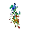
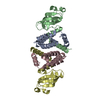
| Deposited unit | 
| ||||||||
|---|---|---|---|---|---|---|---|---|---|
| 1 | 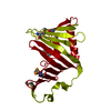
| ||||||||
| Unit cell |
|
-Components
| #1: Protein | Mass: 21820.789 Da / Num. of mol.: 1 Source method: isolated from a genetically manipulated source Source: (gene. exp.) Bacteroides uniformis (bacteria) / Gene: BACUNI_04723 / Plasmid: SpeedET / Production host: Escherichia Coli (E. coli) / Strain (production host): PB1 / References: UniProt: A7VAU1 | ||||
|---|---|---|---|---|---|
| #2: Chemical | Diethylene glycol  Mass: 106.120 Da / Num. of mol.: 2 / Source method: obtained synthetically / Formula: C4H10O3 Mass: 106.120 Da / Num. of mol.: 2 / Source method: obtained synthetically / Formula: C4H10O3#3: Water | ChemComp-HOH / | Water Mass: 18.015 Da / Num. of mol.: 249 / Source method: isolated from a natural source / Formula: H2O Mass: 18.015 Da / Num. of mol.: 249 / Source method: isolated from a natural source / Formula: H2OSequence details | THIS CONSTRUCT (RESIDUES 28-216) WAS EXPRESSED WITH A PURIFICATION TAG MGSDKIHHHHHHENLYFQG. THE TAG ...THIS CONSTRUCT (RESIDUES 28-216) WAS EXPRESSED WITH A PURIFICATI | |
-Experimental details
-Experiment
| Experiment | Method: X-RAY DIFFRACTION / Number of used crystals: 1 |
|---|
- Sample preparation
| Crystal | Density Matthews: 2.24 Å3/Da / Density % sol: 45.21 % |
|---|---|
| Crystal grow | Temperature: 277 K / Method: vapor diffusion, sitting drop Details: 0.20M di-ammonium tartrate, 20.)% polyethylene glycol 3350, NANODROP, VAPOR DIFFUSION, SITTING DROP, temperature 277K |
-Data collection
| Diffraction | Mean temperature: 100 K | |||||||||||||||||||||||||||||||||||||||||||||||||||||||||||||||||||||||||||||
|---|---|---|---|---|---|---|---|---|---|---|---|---|---|---|---|---|---|---|---|---|---|---|---|---|---|---|---|---|---|---|---|---|---|---|---|---|---|---|---|---|---|---|---|---|---|---|---|---|---|---|---|---|---|---|---|---|---|---|---|---|---|---|---|---|---|---|---|---|---|---|---|---|---|---|---|---|---|---|
| Diffraction source | Source: SYNCHROTRON / Site: SSRL  / Beamline: BL11-1 / Wavelength: 0.91837,0.97971,0.97894 / Beamline: BL11-1 / Wavelength: 0.91837,0.97971,0.97894 | |||||||||||||||||||||||||||||||||||||||||||||||||||||||||||||||||||||||||||||
| Detector | Type: DECTRIS PILATUS 6M / Detector: PIXEL / Date: Jul 1, 2013 Details: Flat mirror (vertical focusing); single crystal Si(111) bent monochromator (horizontal focusing) | |||||||||||||||||||||||||||||||||||||||||||||||||||||||||||||||||||||||||||||
| Radiation | Monochromator: single crystal Si(111) bent / Protocol: MAD / Monochromatic (M) / Laue (L): M / Scattering type: x-ray | |||||||||||||||||||||||||||||||||||||||||||||||||||||||||||||||||||||||||||||
| Radiation wavelength |
| |||||||||||||||||||||||||||||||||||||||||||||||||||||||||||||||||||||||||||||
| Reflection | Resolution: 1.4→27.049 Å / Num. obs: 36767 / % possible obs: 89.8 % / Observed criterion σ(I): -3 / Biso Wilson estimate: 15.93 Å2 / Rmerge(I) obs: 0.033 / Net I/σ(I): 10.3 | |||||||||||||||||||||||||||||||||||||||||||||||||||||||||||||||||||||||||||||
| Reflection shell | Diffraction-ID: 1
|
-Phasing
| Phasing | Method: MAD |
|---|
- Processing
| Software |
| ||||||||||||||||||||||||||||||||||||||||||||||||||||||||||||||||||||||||||||||||||||||||||||||||||
|---|---|---|---|---|---|---|---|---|---|---|---|---|---|---|---|---|---|---|---|---|---|---|---|---|---|---|---|---|---|---|---|---|---|---|---|---|---|---|---|---|---|---|---|---|---|---|---|---|---|---|---|---|---|---|---|---|---|---|---|---|---|---|---|---|---|---|---|---|---|---|---|---|---|---|---|---|---|---|---|---|---|---|---|---|---|---|---|---|---|---|---|---|---|---|---|---|---|---|---|
| Refinement | Method to determine structure: MAD / Resolution: 1.4→27.049 Å / Occupancy max: 1 / Occupancy min: 0.12 / SU ML: 0.13 / σ(F): 1.9 / Phase error: 16.44 / Stereochemistry target values: MLHL Details: 1. HYDROGENS HAVE BEEN ADDED IN THE RIDING POSITIONS 2. A MET-INHIBITION PROTOCOL WAS USED FOR SELENOMETHIONINE INCORPORATION DURING PROTEIN EXPRESSION. THE OCCUPANCY OF THE SE ATOMS IN THE ...Details: 1. HYDROGENS HAVE BEEN ADDED IN THE RIDING POSITIONS 2. A MET-INHIBITION PROTOCOL WAS USED FOR SELENOMETHIONINE INCORPORATION DURING PROTEIN EXPRESSION. THE OCCUPANCY OF THE SE ATOMS IN THE MSE RESIDUES WAS REDUCED TO 0.75 TO ACCOUNT FOR THE REDUCED SCATTERING POWER DUE TO PARTIAL S-MET INCORPORATION. 3. ATOM RECORD CONTAINS SUM OF TLS AND RESIDUAL B FACTORS. 4. WATERS WERE EXCLUDED FROM AUTOMATIC TLS ASSIGNMENT. 5.POLYETHYLENE GLYCOL FRAGMENTS (PEG) FROM THE CRYSTALLIZATION HAVE BEEN MODELED INTO THE STRUCTURE.
| ||||||||||||||||||||||||||||||||||||||||||||||||||||||||||||||||||||||||||||||||||||||||||||||||||
| Solvent computation | Shrinkage radii: 0.9 Å / VDW probe radii: 1.11 Å / Solvent model: FLAT BULK SOLVENT MODEL | ||||||||||||||||||||||||||||||||||||||||||||||||||||||||||||||||||||||||||||||||||||||||||||||||||
| Displacement parameters | Biso max: 58.39 Å2 / Biso mean: 20.263 Å2 / Biso min: 8.3 Å2 | ||||||||||||||||||||||||||||||||||||||||||||||||||||||||||||||||||||||||||||||||||||||||||||||||||
| Refinement step | Cycle: LAST / Resolution: 1.4→27.049 Å
| ||||||||||||||||||||||||||||||||||||||||||||||||||||||||||||||||||||||||||||||||||||||||||||||||||
| Refine LS restraints |
| ||||||||||||||||||||||||||||||||||||||||||||||||||||||||||||||||||||||||||||||||||||||||||||||||||
| LS refinement shell | Refine-ID: X-RAY DIFFRACTION / Total num. of bins used: 13
|
