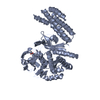
 Movie
Movie Controller


+ Open data

- Basic information
| Entry | Database: PDB / ID: 4mhq | ||||||
|---|---|---|---|---|---|---|---|
| Title | Crystal structure of human primase catalytic subunit | ||||||
 Components Components | DNA primase small subunit Primase Primase | ||||||
 Keywords Keywords | REPLICATION / Zinc finger / Primase / NTP binding / Nucleus | ||||||
| Function / homology |  Function and homology information Function and homology informationDNA primase AEP / ribonucleotide binding / DNA replication initiation / Telomere C-strand synthesis initiation / Inhibition of replication initiation of damaged DNA by RB1/E2F1 / alpha DNA polymerase:primase complex / Polymerase switching / Processive synthesis on the lagging strand / DNA primase activity / Removal of the Flap Intermediate ...DNA primase AEP / ribonucleotide binding / DNA replication initiation / Telomere C-strand synthesis initiation / Inhibition of replication initiation of damaged DNA by RB1/E2F1 / alpha DNA polymerase:primase complex / Polymerase switching / Processive synthesis on the lagging strand / DNA primase activity / Removal of the Flap Intermediate / Polymerase switching on the C-strand of the telomere / DNA replication, synthesis of primer / Activation of the pre-replicative complex / DNA replication initiation / Defective pyroptosis / magnesium ion binding / zinc ion binding / nucleoplasm / membraneSimilarity search - Function | ||||||
| Biological species |  Homo sapiens (human) Homo sapiens (human) | ||||||
| Method | X-RAY DIFFRACTION / SYNCHROTRON / SAD / Resolution: 2.2 Å | ||||||
 Authors Authors | Park, K.R. / An, J.Y. / Lee, Y. / Youn, H.S. / Lee, J.G. / Kang, J.Y. / Kim, T.G. / Lim, J.J. / Eom, S.H. / Wang, J. | ||||||
 Citation Citation | Journal: To be Published Title: Crystal structure of human primase catalytic subunit Authors: Park, K.R. / An, J.Y. / Lee, Y. / Youn, H.S. / Lee, J.G. / Kang, J.Y. / Kim, T.G. / Lim, J.J. / Eom, S.H. / Wang, J. | ||||||
| History |
|
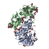
- Structure visualization
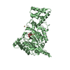
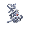
| Structure viewer | Molecule: MolmilJmol/JSmol |
|---|
- Downloads & links
-Download
| PDBx/mmCIF format | 4mhq.cif.gz | 180.4 KB | Display | PDBx/mmCIF format |
|---|---|---|---|---|
| PDB format | pdb4mhq.ent.gz | 143.5 KB | Display | PDB format |
| PDBx/mmJSON format | 4mhq.json.gz | Tree view | PDBx/mmJSON format | |
| Others |  Other downloads Other downloads |
-Validation report
| Arichive directory | https://data.pdbj.org/pub/pdb/validation_reports/mh/4mhqftp://data.pdbj.org/pub/pdb/validation_reports/mh/4mhq | HTTPS FTP |
|---|
-Related structure data
| Similar structure data |
|---|


-Links
 PDBj
PDBj


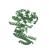
- Assembly
| Deposited unit | 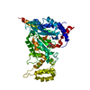
| ||||||||
|---|---|---|---|---|---|---|---|---|---|
| 1 |
| ||||||||
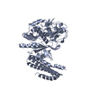
| Unit cell |
|
-Components
| #1: Protein | Primase / DNA primase 49 kDa subunit / p49 Mass: 50384.469 Da / Num. of mol.: 1 / Fragment: Full length Source method: isolated from a genetically manipulated source Source: (gene. exp.) Homo sapiens (human) / Gene: PRIM1 / Plasmid: Modified pET28b / Production host:  Escherichia coli (E. coli) / Strain (production host): BL21(DE3) Escherichia coli (E. coli) / Strain (production host): BL21(DE3)References: UniProt: P49642, Transferases; Transferring phosphorus-containing groups; Nucleotidyltransferases |
|---|---|
| #2: Chemical | ChemComp-ZN /   Mass: 65.409 Da / Num. of mol.: 1 / Source method: obtained synthetically / Formula: Zn Mass: 65.409 Da / Num. of mol.: 1 / Source method: obtained synthetically / Formula: Zn |
| #3: Chemical | ChemComp-CIT / Citric acid  Mass: 192.124 Da / Num. of mol.: 1 / Source method: obtained synthetically / Formula: C6H8O7 Mass: 192.124 Da / Num. of mol.: 1 / Source method: obtained synthetically / Formula: C6H8O7 |
| #4: Water | ChemComp-HOH / Water Mass: 18.015 Da / Num. of mol.: 221 / Source method: isolated from a natural source / Formula: H2O Mass: 18.015 Da / Num. of mol.: 221 / Source method: isolated from a natural source / Formula: H2O |
-Experimental details
-Experiment
| Experiment | Method: X-RAY DIFFRACTION / Number of used crystals: 1 |
|---|
- Sample preparation
| Crystal | Density Matthews: 2.63 Å3/Da / Density % sol: 53.25 % |
|---|---|
| Crystal grow | Temperature: 294 K / Method: vapor diffusion, hanging drop / pH: 4.5 Details: 0.1M Na-acetate, 0.2M Tri-Na-citrate, 20% PEG3350, 0.1M Na-acetate , pH 4.5, VAPOR DIFFUSION, HANGING DROP, temperature 294K |
-Data collection
| Diffraction | Mean temperature: 90 K |
|---|---|
| Diffraction source | Source: SYNCHROTRON / Site: SPring-8  / Beamline: BL26B1 / Wavelength: 1.28262 Å / Beamline: BL26B1 / Wavelength: 1.28262 Å |
| Detector | Type: RIGAKU SATURN A200 / Detector: CCD / Date: Jan 19, 2013 |
| Radiation | Monochromator: Fixed exit Si double crystal monochromator / Protocol: SINGLE WAVELENGTH / Monochromatic (M) / Laue (L): M / Scattering type: x-ray |
| Radiation wavelength | Wavelength: 1.28262 Å / Relative weight: 1 |
| Reflection | Resolution: 2.2→50 Å / Num. all: 27910 / Num. obs: 25240 / % possible obs: 89.6 % / Observed criterion σ(F): 0 / Observed criterion σ(I): 0 / Redundancy: 8.9 % / Rmerge(I) obs: 0.076 / Rsym value: 0.076 / Net I/σ(I): 9.5 |
| Reflection shell | Resolution: 2.2→2.24 Å / Redundancy: 4.7 % / Rmerge(I) obs: 0.976 / Mean I/σ(I) obs: 1.506 / Num. unique all: 1263 / Rsym value: 0.976 / % possible all: 91.5 |
- Processing
| Software |
| |||||||||||||||||||||||||||||||||||||||||||||
|---|---|---|---|---|---|---|---|---|---|---|---|---|---|---|---|---|---|---|---|---|---|---|---|---|---|---|---|---|---|---|---|---|---|---|---|---|---|---|---|---|---|---|---|---|---|---|
| Refinement | Method to determine structure: SAD / Resolution: 2.2→38.06 Å / Cor.coef. Fo:Fc: 0.955 / Cor.coef. Fo:Fc free: 0.922 / SU B: 13.342 / SU ML: 0.165 / Cross valid method: THROUGHOUT / σ(F): 0 / ESU R: 0.307 / ESU R Free: 0.231 / Stereochemistry target values: MAXIMUM LIKELIHOOD
| |||||||||||||||||||||||||||||||||||||||||||||
| Solvent computation | Ion probe radii: 0.8 Å / Shrinkage radii: 0.8 Å / VDW probe radii: 1.2 Å / Solvent model: MASK | |||||||||||||||||||||||||||||||||||||||||||||
| Displacement parameters | Biso mean: 38.961 Å2
| |||||||||||||||||||||||||||||||||||||||||||||
| Refinement step | Cycle: LAST / Resolution: 2.2→38.06 Å
| |||||||||||||||||||||||||||||||||||||||||||||
| Refine LS restraints |
| |||||||||||||||||||||||||||||||||||||||||||||
| LS refinement shell | Resolution: 2.2→2.257 Å / Total num. of bins used: 20
| |||||||||||||||||||||||||||||||||||||||||||||
| Refinement TLS params. | Method: refined / Origin x: 18.7984 Å / Origin y: 26.7721 Å / Origin z: 53.0541 Å
|
