 Movie
Movie Controller

[English] 日本語
 Yorodumi
- PDB-4mh1: Crystal structure and functional studies of quinoprotein L-sorbos... -
+ Open data

- Basic information
| Entry | Database: PDB / ID: 4mh1 | ||||||
|---|---|---|---|---|---|---|---|
| Title | Crystal structure and functional studies of quinoprotein L-sorbose dehydrogenase from Ketogulonicigenium vulgare Y25 | ||||||
 Components Components | Sorbose dehydrogenase | ||||||
 Keywords Keywords | OXIDOREDUCTASE / 2-keto-L-gulonic acid / Ketogulonicigenium vulgare / L-sorbose dehydrogenase / Beta-Propeller / Hydrolase / Carbohydrate/Sugar Binding / periplasmic | ||||||
| Function / homology |  Function and homology information Function and homology informationQuinoprotein alcohol dehydrogenase-like superfamily / Pyrrolo-quinoline quinone repeat / PQQ-like domain / Pyrrolo-quinoline quinone beta-propeller repeat / beta-propeller repeat / 8 Propeller / Methanol Dehydrogenase; Chain A / Quinoprotein alcohol dehydrogenase-like superfamily / Mainly BetaSimilarity search - Domain/homology | ||||||
| Biological species |  Ketogulonicigenium vulgare (bacteria) Ketogulonicigenium vulgare (bacteria) | ||||||
| Method | X-RAY DIFFRACTION / SYNCHROTRON / MOLECULAR REPLACEMENT / Resolution: 2.7 Å | ||||||
 Authors Authors | Han, X. / Liu, X. | ||||||
 Citation Citation | Journal: Biotechnol.Lett. / Year: 2014 Title: Crystal structure of L-sorbose dehydrogenase, a pyrroloquinoline quinone-dependent enzyme with homodimeric assembly, from Ketogulonicigenium vulgare Authors: Han, X. / Xiong, X. / Jiang, D. / Chen, S. / Huang, E. / Zhang, W. / Liu, X. | ||||||
| History |
|
- Structure visualization
| Structure viewer | Molecule: MolmilJmol/JSmol |
|---|
- Downloads & links
-Download
| PDBx/mmCIF format | 4mh1.cif.gz | 386 KB | Display | PDBx/mmCIF format |
|---|---|---|---|---|
| PDB format | pdb4mh1.ent.gz | 315.3 KB | Display | PDB format |
| PDBx/mmJSON format | 4mh1.json.gz | Tree view | PDBx/mmJSON format | |
| Others |  Other downloads Other downloads |
-Validation report
| Arichive directory | https://data.pdbj.org/pub/pdb/validation_reports/mh/4mh1ftp://data.pdbj.org/pub/pdb/validation_reports/mh/4mh1 | HTTPS FTP |
|---|
-Related structure data
| Related structure data |  1yiqS S: Starting model for refinement |
|---|---|
| Similar structure data |


-Links
 PDBj
PDBj
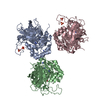
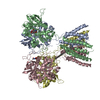
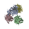
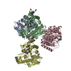
- Assembly
| Deposited unit | 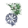
| ||||||||
|---|---|---|---|---|---|---|---|---|---|
| 1 | 
| ||||||||
| Unit cell |
|
-Components
| #1: Protein | / L-sorbose dehydrogenase Mass: 61263.410 Da / Num. of mol.: 2 / Fragment: UNP residues 23-578 / Source method: isolated from a natural source / Source: (natural) Ketogulonicigenium vulgare (bacteria) / Strain: Y25 / References: UniProt: E3F069#2: Chemical | ChemComp-PQQ / | Pyrroloquinoline quinone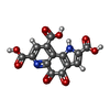  Mass: 330.206 Da / Num. of mol.: 1 / Source method: obtained synthetically / Formula: C14H6N2O8 Mass: 330.206 Da / Num. of mol.: 1 / Source method: obtained synthetically / Formula: C14H6N2O8#3: Chemical | ChemComp-CA / |   Mass: 40.078 Da / Num. of mol.: 1 / Source method: obtained synthetically / Formula: Ca Mass: 40.078 Da / Num. of mol.: 1 / Source method: obtained synthetically / Formula: Ca |
|---|
-Experimental details
-Experiment
| Experiment | Method: X-RAY DIFFRACTION / Number of used crystals: 1 |
|---|
- Sample preparation
| Crystal | Density Matthews: 2.78 Å3/Da / Density % sol: 55.81 % |
|---|---|
| Crystal grow | Temperature: 298 K / Method: vapor diffusion, hanging drop / pH: 7.4 Details: 1.7M ammonium sulfate, 5mM PQQ, 5mM CaCl2, 0.1M HEPES, pH 7.4, VAPOR DIFFUSION, HANGING DROP, temperature 298K |
-Data collection
| Diffraction | Mean temperature: 100 K | ||||||||||||||||||
|---|---|---|---|---|---|---|---|---|---|---|---|---|---|---|---|---|---|---|---|
| Diffraction source | Source: SYNCHROTRON / Site: SSRF  / Beamline: BL17U / Wavelength: 0.9792 Å / Beamline: BL17U / Wavelength: 0.9792 Å | ||||||||||||||||||
| Detector | Type: MAR CCD 165 mm / Detector: CCD / Date: Jun 22, 2012 | ||||||||||||||||||
| Radiation | Monochromator: GRAPHITE / Protocol: SINGLE WAVELENGTH / Monochromatic (M) / Laue (L): M / Scattering type: x-ray | ||||||||||||||||||
| Radiation wavelength | Wavelength: 0.9792 Å / Relative weight: 1 | ||||||||||||||||||
| Reflection | Resolution: 2.7→50 Å / Num. obs: 37049 / % possible obs: 80 % / Observed criterion σ(F): 2 / Observed criterion σ(I): 2 / Biso Wilson estimate: 34.87 Å2 | ||||||||||||||||||
| Reflection shell |
|
- Processing
| Software |
| |||||||||||||||||||||||||||||||||||||||||||||||||||||||||||||||||||||||||||||||||||||||||||||||||||||||||
|---|---|---|---|---|---|---|---|---|---|---|---|---|---|---|---|---|---|---|---|---|---|---|---|---|---|---|---|---|---|---|---|---|---|---|---|---|---|---|---|---|---|---|---|---|---|---|---|---|---|---|---|---|---|---|---|---|---|---|---|---|---|---|---|---|---|---|---|---|---|---|---|---|---|---|---|---|---|---|---|---|---|---|---|---|---|---|---|---|---|---|---|---|---|---|---|---|---|---|---|---|---|---|---|---|---|---|
| Refinement | Method to determine structure: MOLECULAR REPLACEMENT Starting model: 1YiQ Resolution: 2.7→41.137 Å / FOM work R set: 0.8451 / SU ML: 0.32 / σ(F): 1.34 / Phase error: 21.61 / Stereochemistry target values: ML
| |||||||||||||||||||||||||||||||||||||||||||||||||||||||||||||||||||||||||||||||||||||||||||||||||||||||||
| Solvent computation | Shrinkage radii: 0.86 Å / VDW probe radii: 1.1 Å / Solvent model: FLAT BULK SOLVENT MODEL / Bsol: 9.07 Å2 / ksol: 0.328 e/Å3 | |||||||||||||||||||||||||||||||||||||||||||||||||||||||||||||||||||||||||||||||||||||||||||||||||||||||||
| Displacement parameters | Biso max: 141.59 Å2 / Biso mean: 38.69 Å2 / Biso min: 3.4 Å2
| |||||||||||||||||||||||||||||||||||||||||||||||||||||||||||||||||||||||||||||||||||||||||||||||||||||||||
| Refinement step | Cycle: LAST / Resolution: 2.7→41.137 Å
| |||||||||||||||||||||||||||||||||||||||||||||||||||||||||||||||||||||||||||||||||||||||||||||||||||||||||
| Refine LS restraints |
| |||||||||||||||||||||||||||||||||||||||||||||||||||||||||||||||||||||||||||||||||||||||||||||||||||||||||
| LS refinement shell | Refine-ID: X-RAY DIFFRACTION / Total num. of bins used: 14
| |||||||||||||||||||||||||||||||||||||||||||||||||||||||||||||||||||||||||||||||||||||||||||||||||||||||||
| Refinement TLS params. | Method: refined / Origin x: 40.8943 Å / Origin y: -32.0454 Å / Origin z: -77.2712 Å
| |||||||||||||||||||||||||||||||||||||||||||||||||||||||||||||||||||||||||||||||||||||||||||||||||||||||||
| Refinement TLS group | Selection details: ALL |
