 Movie
Movie Controller


+ Open data

- Basic information
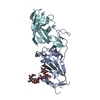
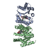
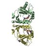
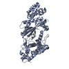
| Entry | Database: PDB / ID: 4mew | ||||||
|---|---|---|---|---|---|---|---|
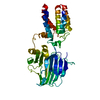
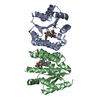
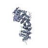
| Title | Structure of the core fragment of human PR70 | ||||||
 Components Components | Serine/threonine-protein phosphatase 2A regulatory subunit B'' subunit beta | ||||||
 Keywords Keywords |  hydrolase / cell cycle / EF-hands / Protein Phosphatase / Calcium Binding / METAL BINDING PROTEIN hydrolase / cell cycle / EF-hands / Protein Phosphatase / Calcium Binding / METAL BINDING PROTEIN | ||||||
| Function / homology |  Function and homology information Function and homology informationprotein phosphatase type 2A complex / Inhibition of replication initiation of damaged DNA by RB1/E2F1 / protein phosphatase regulator activity / Cyclin A/B1/B2 associated events during G2/M transition / protein dephosphorylation / Cyclin D associated events in G1 / regulation of cell cycle / calcium ion binding / nucleoplasm / nucleus / cytosolSimilarity search - Function | ||||||
| Biological species |  Homo sapiens (human) Homo sapiens (human) | ||||||
| Method | X-RAY DIFFRACTION / SYNCHROTRON / SAD / Resolution: 1.993 Å | ||||||
 Authors Authors | Dovega, R.B. / Quistgaard, E.M. / Tsutakawa, S. / Anandapadamanaban, M. / Low, C. / Nordlund, P. | ||||||
 Citation Citation | Journal: Plos One / Year: 2014 Title: Structural and Biochemical Characterization of Human PR70 in Isolation and in Complex with the Scaffolding Subunit of Protein Phosphatase 2A. Authors: Dovega, R. / Tsutakawa, S. / Quistgaard, E.M. / Anandapadamanaban, M. / Low, C. / Nordlund, P. | ||||||
| History |
|
- Structure visualization
| Structure viewer | Molecule: MolmilJmol/JSmol |
|---|
- Downloads & links
-Download
| PDBx/mmCIF format | 4mew.cif.gz | 158.5 KB | Display | PDBx/mmCIF format |
|---|---|---|---|---|
| PDB format | pdb4mew.ent.gz | 130.8 KB | Display | PDB format |
| PDBx/mmJSON format | 4mew.json.gz | Tree view | PDBx/mmJSON format | |
| Others |  Other downloads Other downloads |
-Validation report
| Arichive directory | https://data.pdbj.org/pub/pdb/validation_reports/me/4mewftp://data.pdbj.org/pub/pdb/validation_reports/me/4mew | HTTPS FTP |
|---|
-Related structure data
| Similar structure data |
|---|


-Links
 PDBj
PDBj


- Assembly
| Deposited unit | 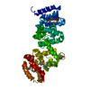
| ||||||||
|---|---|---|---|---|---|---|---|---|---|
| 1 |
| ||||||||
| Unit cell |
|
-Components
| #1: Protein | Mass: 41866.066 Da / Num. of mol.: 1 / Fragment: Residues 130-483 Source method: isolated from a genetically manipulated source Source: (gene. exp.) Homo sapiens (human) / Gene: PPP2R3B, PPP2R3L / Plasmid: pNIC28-Bsa4 / Production host:  Escherichia coli (E. coli) / Strain (production host): Bl21(DE3) / References: UniProt: Q9Y5P8 Escherichia coli (E. coli) / Strain (production host): Bl21(DE3) / References: UniProt: Q9Y5P8 | ||||
|---|---|---|---|---|---|
| #2: Chemical |   Mass: 40.078 Da / Num. of mol.: 2 / Source method: obtained synthetically / Formula: Ca Mass: 40.078 Da / Num. of mol.: 2 / Source method: obtained synthetically / Formula: Ca#3: Chemical | Glycerol  Mass: 92.094 Da / Num. of mol.: 2 / Source method: obtained synthetically / Formula: C3H8O3 Mass: 92.094 Da / Num. of mol.: 2 / Source method: obtained synthetically / Formula: C3H8O3#4: Water | ChemComp-HOH / | Water Mass: 18.015 Da / Num. of mol.: 347 / Source method: isolated from a natural source / Formula: H2O Mass: 18.015 Da / Num. of mol.: 347 / Source method: isolated from a natural source / Formula: H2O |
-Experimental details
-Experiment
| Experiment | Method: X-RAY DIFFRACTION / Number of used crystals: 1 |
|---|
- Sample preparation
| Crystal | Density Matthews: 2.05 Å3/Da / Density % sol: 40.14 % |
|---|---|
| Crystal grow | Temperature: 277 K / Method: vapor diffusion / pH: 7.5 Details: 0.1 M Tris-HCl, 16% PEG 2000 monomethyl ether, 0.15 M trimethylamine N-oxide, pH 7.5, VAPOR DIFFUSION, temperature 277K |
-Data collection
| Diffraction | Mean temperature: 100 K |
|---|---|
| Diffraction source | Source: SYNCHROTRON / Site: Diamond  / Beamline: I04 / Wavelength: 0.9796 Å / Beamline: I04 / Wavelength: 0.9796 Å |
| Detector | Type: ADSC QUANTUM 315 / Detector: CCD / Date: Mar 22, 2012 |
| Radiation | Monochromator: Double crystal, Si(111) / Protocol: SINGLE WAVELENGTH / Monochromatic (M) / Laue (L): M / Scattering type: x-ray |
| Radiation wavelength | Wavelength: 0.9796 Å / Relative weight: 1 |
| Reflection | Resolution: 1.99→37.23 Å / Num. all: 45116 / Num. obs: 45116 / % possible obs: 99.1 % / Observed criterion σ(F): 2 / Observed criterion σ(I): 2 / Rsym value: 0.097 / Net I/σ(I): 13.8 |
| Reflection shell | Resolution: 1.99→2.04 Å / Mean I/σ(I) obs: 2.42 / Num. unique all: 3010 / Rsym value: 0.601 / % possible all: 89 |
- Processing
| Software |
| ||||||||||||||||||||||||||||||||||||||||||||||||||||||||||||||||||||||||||||||||||||||||||||||||||
|---|---|---|---|---|---|---|---|---|---|---|---|---|---|---|---|---|---|---|---|---|---|---|---|---|---|---|---|---|---|---|---|---|---|---|---|---|---|---|---|---|---|---|---|---|---|---|---|---|---|---|---|---|---|---|---|---|---|---|---|---|---|---|---|---|---|---|---|---|---|---|---|---|---|---|---|---|---|---|---|---|---|---|---|---|---|---|---|---|---|---|---|---|---|---|---|---|---|---|---|
| Refinement | Method to determine structure: SAD / Resolution: 1.993→37.23 Å / SU ML: 0.2 / σ(F): 1.51 / Phase error: 20.63 / Stereochemistry target values: ML
| ||||||||||||||||||||||||||||||||||||||||||||||||||||||||||||||||||||||||||||||||||||||||||||||||||
| Solvent computation | Shrinkage radii: 0.9 Å / VDW probe radii: 1.11 Å / Solvent model: FLAT BULK SOLVENT MODEL | ||||||||||||||||||||||||||||||||||||||||||||||||||||||||||||||||||||||||||||||||||||||||||||||||||
| Refinement step | Cycle: LAST / Resolution: 1.993→37.23 Å
| ||||||||||||||||||||||||||||||||||||||||||||||||||||||||||||||||||||||||||||||||||||||||||||||||||
| Refine LS restraints |
| ||||||||||||||||||||||||||||||||||||||||||||||||||||||||||||||||||||||||||||||||||||||||||||||||||
| LS refinement shell |
| ||||||||||||||||||||||||||||||||||||||||||||||||||||||||||||||||||||||||||||||||||||||||||||||||||
| Refinement TLS params. | Method: refined / Refine-ID: X-RAY DIFFRACTION
| ||||||||||||||||||||||||||||||||||||||||||||||||||||||||||||||||||||||||||||||||||||||||||||||||||
| Refinement TLS group |
|
