 Movie
Movie Controller


+ Open data

- Basic information
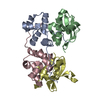
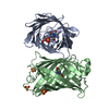
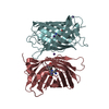
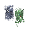
| Entry | Database: PDB / ID: 4mct | ||||||
|---|---|---|---|---|---|---|---|
| Title | P. vulgaris HIGBA structure, crystal form 1 | ||||||
 Components Components |
| ||||||
 Keywords Keywords |  TOXIN / bacterial toxins / biofilms / cell metabolism / energy metabolism / helix-turn-helix transcription factors / microbial pathogenesis / stress response / stringent response / transcription repressor / translation control TOXIN / bacterial toxins / biofilms / cell metabolism / energy metabolism / helix-turn-helix transcription factors / microbial pathogenesis / stress response / stringent response / transcription repressor / translation control | ||||||
| Function / homology |  Function and homology information Function and homology informationtoxin sequestering activity / plasmid maintenance / RNA catabolic process / translation repressor activity / RNA endonuclease activity / negative regulation of cell growth / ribosome binding / Hydrolases; Acting on ester bonds / negative regulation of translation / transcription cis-regulatory region binding ...toxin sequestering activity / plasmid maintenance / RNA catabolic process / translation repressor activity / RNA endonuclease activity / negative regulation of cell growth / ribosome binding / Hydrolases; Acting on ester bonds / negative regulation of translation / transcription cis-regulatory region binding / protein heterodimerization activity / DNA-binding transcription factor activity / negative regulation of cell population proliferation / mRNA binding / regulation of DNA-templated transcriptionSimilarity search - Function | ||||||
| Biological species |  Proteus vulgaris (bacteria) Proteus vulgaris (bacteria) | ||||||
| Method | X-RAY DIFFRACTION / SYNCHROTRON / SAD / Resolution: 2.8 Å | ||||||
 Authors Authors | Schureck, M.A. / Maehigashi, T. / Dunham, C.M. | ||||||
 Citation Citation | Journal: J.Biol.Chem. / Year: 2014 Title: Structure of the Proteus vulgaris HigB-(HigA)2-HigB Toxin-Antitoxin Complex. Authors: Schureck, M.A. / Maehigashi, T. / Miles, S.J. / Marquez, J. / Cho, S.E. / Erdman, R. / Dunham, C.M. | ||||||
| History |
|
- Structure visualization
| Structure viewer | Molecule: MolmilJmol/JSmol |
|---|
- Downloads & links
-Download
| PDBx/mmCIF format | 4mct.cif.gz | 82.2 KB | Display | PDBx/mmCIF format |
|---|---|---|---|---|
| PDB format | pdb4mct.ent.gz | 66.5 KB | Display | PDB format |
| PDBx/mmJSON format | 4mct.json.gz | Tree view | PDBx/mmJSON format | |
| Others |  Other downloads Other downloads |
-Validation report
| Arichive directory | https://data.pdbj.org/pub/pdb/validation_reports/mc/4mctftp://data.pdbj.org/pub/pdb/validation_reports/mc/4mct | HTTPS FTP |
|---|
-Related structure data


-Links
 PDBj
PDBj


- Assembly
| Deposited unit | 
| ||||||||
|---|---|---|---|---|---|---|---|---|---|
| 1 |
| ||||||||
| Unit cell |
| ||||||||
| Components on special symmetry positions |
|
-Components
| #1: Protein | / Host inhibition of growth A Mass: 14019.115 Da / Num. of mol.: 2 Source method: isolated from a genetically manipulated source Source: (gene. exp.) Proteus vulgaris (bacteria) / Strain: UR-75 / Gene: higA / Plasmid: pET21c / Production host: Escherichia coli (E. coli) / Strain (production host): BL21(DE3) / References: UniProt: Q7A224#2: Protein | / Host inhibition of growth BMass: 10983.193 Da / Num. of mol.: 2 Source method: isolated from a genetically manipulated source Source: (gene. exp.) Proteus vulgaris (bacteria) / Strain: UR-75 / Gene: higB / Plasmid: pET21c / Production host: Escherichia coli (E. coli) / Strain (production host): BL21(DE3) / References: UniProt: Q7A225#3: Water | ChemComp-HOH / | Water Mass: 18.015 Da / Num. of mol.: 40 / Source method: isolated from a natural source / Formula: H2O Mass: 18.015 Da / Num. of mol.: 40 / Source method: isolated from a natural source / Formula: H2O |
|---|
-Experimental details
-Experiment
| Experiment | Method: X-RAY DIFFRACTION / Number of used crystals: 1 |
|---|
- Sample preparation
| Crystal | Density Matthews: 3.37 Å3/Da / Density % sol: 63.46 % |
|---|---|
| Crystal grow | Temperature: 293.15 K / pH: 7.5 Details: 3-10 % PEG 3350, 0.2 M L-proline, 0.1 M HEPES pH 7.5, VAPOR DIFFUSION, SITTING DROP, temperature 293.15K |
-Data collection
| Diffraction | Mean temperature: 100 K | |||||||||||||||||||||||||||||||||||||||||||||||||||||||||||||||||||||||||||||||||||||||||||||||||||||||||||||||||||||||||||||||||||||||||||||||||||
|---|---|---|---|---|---|---|---|---|---|---|---|---|---|---|---|---|---|---|---|---|---|---|---|---|---|---|---|---|---|---|---|---|---|---|---|---|---|---|---|---|---|---|---|---|---|---|---|---|---|---|---|---|---|---|---|---|---|---|---|---|---|---|---|---|---|---|---|---|---|---|---|---|---|---|---|---|---|---|---|---|---|---|---|---|---|---|---|---|---|---|---|---|---|---|---|---|---|---|---|---|---|---|---|---|---|---|---|---|---|---|---|---|---|---|---|---|---|---|---|---|---|---|---|---|---|---|---|---|---|---|---|---|---|---|---|---|---|---|---|---|---|---|---|---|---|---|---|---|
| Diffraction source | Source: SYNCHROTRON / Site: APS  / Beamline: 24-ID-C / Wavelength: 0.97922 / Beamline: 24-ID-C / Wavelength: 0.97922 | |||||||||||||||||||||||||||||||||||||||||||||||||||||||||||||||||||||||||||||||||||||||||||||||||||||||||||||||||||||||||||||||||||||||||||||||||||
| Detector | Type: ADSC QUANTUM 315 / Detector: CCD / Date: Dec 8, 2011 / Details: MIRRORS | |||||||||||||||||||||||||||||||||||||||||||||||||||||||||||||||||||||||||||||||||||||||||||||||||||||||||||||||||||||||||||||||||||||||||||||||||||
| Radiation | Monochromator: KOHZU DIAMOND MONOCHROMATOR / Protocol: SINGLE WAVELENGTH / Monochromatic (M) / Laue (L): M / Scattering type: x-ray | |||||||||||||||||||||||||||||||||||||||||||||||||||||||||||||||||||||||||||||||||||||||||||||||||||||||||||||||||||||||||||||||||||||||||||||||||||
| Radiation wavelength | Wavelength: 0.97922 Å / Relative weight: 1 | |||||||||||||||||||||||||||||||||||||||||||||||||||||||||||||||||||||||||||||||||||||||||||||||||||||||||||||||||||||||||||||||||||||||||||||||||||
| Reflection | Redundancy: 6.6 % / Number: 125944 / Rmerge(I) obs: 0.163 / Χ2: 1.81 / D res high: 2.7 Å / D res low: 40 Å / Num. obs: 18970 / % possible obs: 100 | |||||||||||||||||||||||||||||||||||||||||||||||||||||||||||||||||||||||||||||||||||||||||||||||||||||||||||||||||||||||||||||||||||||||||||||||||||
| Diffraction reflection shell |
| |||||||||||||||||||||||||||||||||||||||||||||||||||||||||||||||||||||||||||||||||||||||||||||||||||||||||||||||||||||||||||||||||||||||||||||||||||
| Reflection | Resolution: 2.8→40 Å / Num. obs: 35400 / % possible obs: 100 % / Observed criterion σ(I): -3 / Redundancy: 6.8 % / Biso Wilson estimate: 63.46 Å2 / Rmerge(I) obs: 0.155 / Net I/σ(I): 13.8 | |||||||||||||||||||||||||||||||||||||||||||||||||||||||||||||||||||||||||||||||||||||||||||||||||||||||||||||||||||||||||||||||||||||||||||||||||||
| Reflection shell | Resolution: 2.8→2.9 Å / Redundancy: 6.2 % / Rmerge(I) obs: 0.752 / Mean I/σ(I) obs: 2.1 / % possible all: 97.2 |
- Processing
| Software |
| |||||||||||||||||||||||||||||||||||||||||||||||||||||||||||||||||||||||||||||||||||||||||||||||||||||||||||||||||||||||||||||||||||||||||||||||||||||||||||||||||
|---|---|---|---|---|---|---|---|---|---|---|---|---|---|---|---|---|---|---|---|---|---|---|---|---|---|---|---|---|---|---|---|---|---|---|---|---|---|---|---|---|---|---|---|---|---|---|---|---|---|---|---|---|---|---|---|---|---|---|---|---|---|---|---|---|---|---|---|---|---|---|---|---|---|---|---|---|---|---|---|---|---|---|---|---|---|---|---|---|---|---|---|---|---|---|---|---|---|---|---|---|---|---|---|---|---|---|---|---|---|---|---|---|---|---|---|---|---|---|---|---|---|---|---|---|---|---|---|---|---|---|---|---|---|---|---|---|---|---|---|---|---|---|---|---|---|---|---|---|---|---|---|---|---|---|---|---|---|---|---|---|---|---|
| Refinement | Method to determine structure: SAD / Resolution: 2.8→41.08 Å / Occupancy max: 1 / Occupancy min: 1 / SU ML: 0.39 / σ(F): 1.34 / Phase error: 24.95 / Stereochemistry target values: ML
| |||||||||||||||||||||||||||||||||||||||||||||||||||||||||||||||||||||||||||||||||||||||||||||||||||||||||||||||||||||||||||||||||||||||||||||||||||||||||||||||||
| Solvent computation | Shrinkage radii: 0.9 Å / VDW probe radii: 1.11 Å / Solvent model: FLAT BULK SOLVENT MODEL | |||||||||||||||||||||||||||||||||||||||||||||||||||||||||||||||||||||||||||||||||||||||||||||||||||||||||||||||||||||||||||||||||||||||||||||||||||||||||||||||||
| Displacement parameters | Biso mean: 40.74 Å2 | |||||||||||||||||||||||||||||||||||||||||||||||||||||||||||||||||||||||||||||||||||||||||||||||||||||||||||||||||||||||||||||||||||||||||||||||||||||||||||||||||
| Refinement step | Cycle: LAST / Resolution: 2.8→41.08 Å
| |||||||||||||||||||||||||||||||||||||||||||||||||||||||||||||||||||||||||||||||||||||||||||||||||||||||||||||||||||||||||||||||||||||||||||||||||||||||||||||||||
| Refine LS restraints |
| |||||||||||||||||||||||||||||||||||||||||||||||||||||||||||||||||||||||||||||||||||||||||||||||||||||||||||||||||||||||||||||||||||||||||||||||||||||||||||||||||
| LS refinement shell |
|
