 Movie
Movie Controller

[English] 日本語
 Yorodumi
- PDB-4m7r: Crystal structure of the N-terminal methyltransferase-like domain... -
+ Open data

- Basic information
| Entry | Database: PDB / ID: 4m7r | ||||||
|---|---|---|---|---|---|---|---|
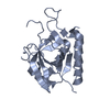
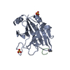
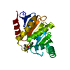
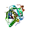
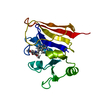
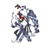
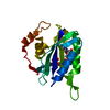
| Title | Crystal structure of the N-terminal methyltransferase-like domain of anamorsin | ||||||
 Components Components | Anamorsin | ||||||
 Keywords Keywords |  APOPTOSIS / ROSSMANN FOLD APOPTOSIS / ROSSMANN FOLD | ||||||
| Function / homology |  Function and homology information Function and homology informationCytosolic iron-sulfur cluster assembly / iron-sulfur cluster assembly / hemopoiesis / mitochondrial intermembrane space / 2 iron, 2 sulfur cluster binding / 4 iron, 4 sulfur cluster binding / electron transfer activity / iron ion binding / apoptotic process / nucleolus ...Cytosolic iron-sulfur cluster assembly / iron-sulfur cluster assembly / hemopoiesis / mitochondrial intermembrane space / 2 iron, 2 sulfur cluster binding / 4 iron, 4 sulfur cluster binding / electron transfer activity / iron ion binding / apoptotic process / nucleolus / negative regulation of apoptotic process / mitochondrion / nucleoplasm / cytoplasmSimilarity search - Function | ||||||
| Biological species |  Homo sapiens (human) Homo sapiens (human) | ||||||
| Method | X-RAY DIFFRACTION / SYNCHROTRON / SAD / Resolution: 1.801 Å | ||||||
 Authors Authors | Song, G. / Liu, Z.-J. | ||||||
 Citation Citation | Journal: Proteins / Year: 2014 Title: Crystal structure of the N-terminal methyltransferase-like domain of anamorsin. Authors: Song, G. / Cheng, C. / Li, Y. / Shaw, N. / Xiao, Z.C. / Liu, Z.J. | ||||||
| History |
|
- Structure visualization
| Structure viewer | Molecule: MolmilJmol/JSmol |
|---|
- Downloads & links
-Download
| PDBx/mmCIF format | 4m7r.cif.gz | 157.2 KB | Display | PDBx/mmCIF format |
|---|---|---|---|---|
| PDB format | pdb4m7r.ent.gz | 125.1 KB | Display | PDB format |
| PDBx/mmJSON format | 4m7r.json.gz | Tree view | PDBx/mmJSON format | |
| Others |  Other downloads Other downloads |
-Validation report
| Arichive directory | https://data.pdbj.org/pub/pdb/validation_reports/m7/4m7rftp://data.pdbj.org/pub/pdb/validation_reports/m7/4m7r | HTTPS FTP |
|---|
-Related structure data
| Similar structure data |
|---|


-Links
 PDBj
PDBj


- Assembly
| Deposited unit | 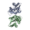
| ||||||||
|---|---|---|---|---|---|---|---|---|---|
| 1 | 
| ||||||||
| 2 | 
| ||||||||
| Unit cell |
|
-Components
| #1: Protein | Mass: 21283.066 Da / Num. of mol.: 2 / Fragment: methyltransferase-like domain, UNP residues 1-172 Source method: isolated from a genetically manipulated source Source: (gene. exp.) Homo sapiens (human) / Gene: CIAPIN1, CUA001, PRO0915 / Plasmid: PMCSG7 / Production host:  Escherichia coli (E. coli) / Strain (production host): BL21 / References: UniProt: Q6FI81 Escherichia coli (E. coli) / Strain (production host): BL21 / References: UniProt: Q6FI81#2: Chemical | ChemComp-HG / | Mercury (element)  Mass: 200.590 Da / Num. of mol.: 1 / Source method: obtained synthetically / Formula: Hg Mass: 200.590 Da / Num. of mol.: 1 / Source method: obtained synthetically / Formula: Hg#3: Water | ChemComp-HOH / | Water Mass: 18.015 Da / Num. of mol.: 281 / Source method: isolated from a natural source / Formula: H2O Mass: 18.015 Da / Num. of mol.: 281 / Source method: isolated from a natural source / Formula: H2O |
|---|
-Experimental details
-Experiment
| Experiment | Method: X-RAY DIFFRACTION / Number of used crystals: 2 |
|---|
- Sample preparation
| Crystal | Density Matthews: 2.18 Å3/Da / Density % sol: 43.61 % |
|---|---|
| Crystal grow | Temperature: 289 K / Method: evaporation / pH: 6.5 Details: 0.17M sodium acetate trihydrate, 0.085M sodium cacodylate pH6.5, 25% PEG 8000, 15% Glycerol, EVAPORATION, temperature 289K |
-Data collection
| Diffraction | Mean temperature: 100 K | ||||||||||||||||||||||||||||||||||||||||||
|---|---|---|---|---|---|---|---|---|---|---|---|---|---|---|---|---|---|---|---|---|---|---|---|---|---|---|---|---|---|---|---|---|---|---|---|---|---|---|---|---|---|---|---|
| Diffraction source | Source: SYNCHROTRON / Site: APS  / Beamline: 19-ID / Wavelength: 1 Å / Beamline: 19-ID / Wavelength: 1 Å | ||||||||||||||||||||||||||||||||||||||||||
| Detector | Type: ADSC QUANTUM 315r / Detector: CCD / Date: Jun 14, 2008 | ||||||||||||||||||||||||||||||||||||||||||
| Radiation | Monochromator: Rosenbaum-Rock high-resolution double-crystal monochromatoR Protocol: SINGLE WAVELENGTH / Monochromatic (M) / Laue (L): M / Scattering type: x-ray | ||||||||||||||||||||||||||||||||||||||||||
| Radiation wavelength | Wavelength: 1 Å / Relative weight: 1 | ||||||||||||||||||||||||||||||||||||||||||
| Reflection | Resolution: 1.8→30.43 Å / Num. all: 33762 / Num. obs: 33762 / % possible obs: 100 % / Observed criterion σ(F): 1 / Observed criterion σ(I): 1 / Redundancy: 10.7 % / Biso Wilson estimate: 21.95 Å2 / Rmerge(I) obs: 0.067 / Rsym value: 0.067 / Net I/σ(I): 42.9 | ||||||||||||||||||||||||||||||||||||||||||
| Reflection shell | Diffraction-ID: 1 / % possible all: 100
|
- Processing
| Software |
| ||||||||||||||||||||||||||||||||||||||||||||||||||||||||||||||||||||||||||||||
|---|---|---|---|---|---|---|---|---|---|---|---|---|---|---|---|---|---|---|---|---|---|---|---|---|---|---|---|---|---|---|---|---|---|---|---|---|---|---|---|---|---|---|---|---|---|---|---|---|---|---|---|---|---|---|---|---|---|---|---|---|---|---|---|---|---|---|---|---|---|---|---|---|---|---|---|---|---|---|---|
| Refinement | Method to determine structure: SAD / Resolution: 1.801→30.426 Å / Occupancy max: 1 / Occupancy min: 0.3 / FOM work R set: 0.8364 / SU ML: 0.2 / σ(F): 1.33 / Phase error: 23.71 / Stereochemistry target values: ML
| ||||||||||||||||||||||||||||||||||||||||||||||||||||||||||||||||||||||||||||||
| Solvent computation | Shrinkage radii: 0.9 Å / VDW probe radii: 1.11 Å / Solvent model: FLAT BULK SOLVENT MODEL | ||||||||||||||||||||||||||||||||||||||||||||||||||||||||||||||||||||||||||||||
| Displacement parameters | Biso max: 72.73 Å2 / Biso mean: 28.2786 Å2 / Biso min: 10.38 Å2 | ||||||||||||||||||||||||||||||||||||||||||||||||||||||||||||||||||||||||||||||
| Refinement step | Cycle: LAST / Resolution: 1.801→30.426 Å
| ||||||||||||||||||||||||||||||||||||||||||||||||||||||||||||||||||||||||||||||
| Refine LS restraints |
| ||||||||||||||||||||||||||||||||||||||||||||||||||||||||||||||||||||||||||||||
| LS refinement shell | Refine-ID: X-RAY DIFFRACTION / Total num. of bins used: 12 / % reflection obs: 100 %
| ||||||||||||||||||||||||||||||||||||||||||||||||||||||||||||||||||||||||||||||
| Refinement TLS params. | Method: refined / Origin x: 39.2964 Å / Origin y: -13.8878 Å / Origin z: -17.3673 Å
| ||||||||||||||||||||||||||||||||||||||||||||||||||||||||||||||||||||||||||||||
| Refinement TLS group |
|
