 Movie
Movie Controller


+ Open data

- Basic information
| Entry | Database: PDB / ID: 4m6f | ||||||
|---|---|---|---|---|---|---|---|
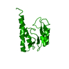
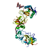
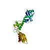
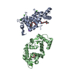
| Title | Dimer of the G-Segment Invertase bound to a DNA substrate | ||||||
 Components Components |
| ||||||
 Keywords Keywords | HYDROLASE/DNA /  Helix-turn-helix / catalytic domain / DNA Binding Domain / recombinase / HYDROLASE-DNA complex Helix-turn-helix / catalytic domain / DNA Binding Domain / recombinase / HYDROLASE-DNA complex | ||||||
| Function / homology |  Function and homology information Function and homology informationviral tropism switching / Ligases; Forming phosphoric-ester bonds / Hydrolases; Acting on ester bonds; Endodeoxyribonucleases producing 3'-phosphomonoesters / DNA strand exchange activity / ligase activity / DNA integration / host cell cytoplasm / hydrolase activity / symbiont entry into host cell / DNA bindingSimilarity search - Function | ||||||
| Biological species |  Enterobacteria phage Mu (virus) Enterobacteria phage Mu (virus)synthetic construct (others) | ||||||
| Method | X-RAY DIFFRACTION / SYNCHROTRON / SAD / Resolution: 4.99 Å | ||||||
 Authors Authors | Ritacco, C.J. / Wang, J. / Steitz, T.A. | ||||||
 Citation Citation | Journal: Acta Crystallogr.,Sect.D / Year: 2014 Title: Exploiting large non-isomorphous differences for phase determination of a G-segment invertase-DNA complex. Authors: Ritacco, C.J. / Steitz, T.A. / Wang, J. | ||||||
| History |
|
- Structure visualization
| Structure viewer | Molecule: MolmilJmol/JSmol |
|---|
- Downloads & links
-Download
| PDBx/mmCIF format | 4m6f.cif.gz | 65 KB | Display | PDBx/mmCIF format |
|---|---|---|---|---|
| PDB format | pdb4m6f.ent.gz | 48.3 KB | Display | PDB format |
| PDBx/mmJSON format | 4m6f.json.gz | Tree view | PDBx/mmJSON format | |
| Others |  Other downloads Other downloads |
-Validation report
| Arichive directory | https://data.pdbj.org/pub/pdb/validation_reports/m6/4m6fftp://data.pdbj.org/pub/pdb/validation_reports/m6/4m6f | HTTPS FTP |
|---|
-Related structure data
| Related structure data | |
|---|---|
| Similar structure data |


-Links
 PDBj
PDBj


- Assembly
| Deposited unit | 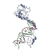
| ||||||||
|---|---|---|---|---|---|---|---|---|---|
| 1 |
| ||||||||
| Unit cell |
|
-Components
| #1: Protein | Mass: 21714.197 Da / Num. of mol.: 1 / Source method: isolated from a natural source / Source: (natural) Enterobacteria phage Mu (virus) / References: UniProt: P03015 |
|---|---|
| #2: DNA chain | Mass: 5246.429 Da / Num. of mol.: 1 / Source method: obtained synthetically / Source: (synth.) synthetic construct (others) |
| #3: DNA chain | Mass: 5475.620 Da / Num. of mol.: 1 / Source method: obtained synthetically / Source: (synth.) synthetic construct (others) |
-Experimental details
-Experiment
| Experiment | Method: X-RAY DIFFRACTION / Number of used crystals: 3 |
|---|
- Sample preparation
| Crystal grow | Temperature: 289 K / Method: vapor diffusion, hanging drop / pH: 6.5 Details: 3-5% Ammonium Sulfate 20% ethylene glycol 100 mM MES, pH 6.5, VAPOR DIFFUSION, HANGING DROP, temperature 289K |
|---|
-Data collection
| Diffraction |
| ||||||||||||||||||||||||
|---|---|---|---|---|---|---|---|---|---|---|---|---|---|---|---|---|---|---|---|---|---|---|---|---|---|
| Diffraction source |
| ||||||||||||||||||||||||
| Detector |
| ||||||||||||||||||||||||
| Radiation |
| ||||||||||||||||||||||||
| Radiation wavelength |
| ||||||||||||||||||||||||
| Reflection | Resolution: 4.99→30 Å / % possible obs: 94.5 % / Observed criterion σ(F): 1 / Observed criterion σ(I): 1 / Redundancy: 12.5 % / Rmerge(I) obs: 0.111 / Rsym value: 12.05 | ||||||||||||||||||||||||
| Reflection shell | Resolution: 4.97→5.25 Å / % possible all: 94.6 |

- Processing
| Software |
| ||||||||||||||||||
|---|---|---|---|---|---|---|---|---|---|---|---|---|---|---|---|---|---|---|---|
| Refinement | Method to determine structure: SAD / Resolution: 4.99→30 Å / σ(F): 1 / Stereochemistry target values: DEN
| ||||||||||||||||||
| Displacement parameters | Biso mean: 335 Å2
| ||||||||||||||||||
| Refinement step | Cycle: LAST / Resolution: 4.99→30 Å
|
