Mass: 18.015 Da / Num. of mol.: 225 / Source method: isolated from a natural source / Formula: H2O
-
Experimental details
-
Experiment
Experiment
Method: X-RAY DIFFRACTION
-
Sample preparation
Crystal
Density Matthews: 2.6 Å3/Da / Density % sol: 52.63 %
Crystal grow
Temperature: 293 K / Method: vapor diffusion, sitting drop / pH: 6.7 Details: 200 MM MES, 27% PEG8K, 200 MM AMMONIUM SULFATE, 1 MM MANGANESE CHLORIDE, 10 MM MAGNESIUM ACETATE, 10 MM TAURINE, AND 50 MM SODIUM FLUORIDE, pH 6.7, VAPOR DIFFUSION, SITTING DROP, temperature 293K
Monochromator: HORIZONTAL BENT SI(111), ASYMMETRICALLY CUT WITH WATER COOLED CU BLOCK Protocol: SINGLE WAVELENGTH / Monochromatic (M) / Laue (L): M / Scattering type: x-ray
Radiation wavelength
Wavelength: 1.1 Å / Relative weight: 1
Reflection
Resolution: 1.53→50.75 Å / Num. obs: 250493 / % possible obs: 98 % / Observed criterion σ(I): -3 / Redundancy: 5.6 % / Rmerge(I) obs: 0.041 / Net I/σ(I): 16.2
Reflection shell
Resolution: 1.53→1.62 Å / Redundancy: 4.6 % / Rmerge(I) obs: 0.369 / Mean I/σ(I) obs: 3.2 / % possible all: 97.7
-
Processing
Software
Name
Version
Classification
CBASS
datacollection
PHENIX
(PHENIX.REFINE: 1.8.2_1309)
modelbuilding
PHENIX
(phenix.refine: 1.8.2_1309)
refinement
MOSFLM
datareduction
SCALA
datascaling
PHENIX
1.8.2_1309
phasing
Refinement
Method to determine structure: MOLECULAR REPLACEMENT / Resolution: 1.534→46.9 Å / SU ML: 0.1 / σ(F): 1.34 / Phase error: 20.76 / Stereochemistry target values: ML
Rfactor
Num. reflection
% reflection
Rfree
0.1839
889
2 %
Rwork
0.1707
-
-
obs
0.1709
44527
99.52 %
Solvent computation
Shrinkage radii: 0.9 Å / VDW probe radii: 1.11 Å / Solvent model: FLAT BULK SOLVENT MODEL
Refinement step
Cycle: LAST / Resolution: 1.534→46.9 Å
Protein
Nucleic acid
Ligand
Solvent
Total
Num. atoms
1657
0
7
225
1889
Refine LS restraints
Refine-ID
Type
Dev ideal
Number
X-RAY DIFFRACTION
f_bond_d
0.013
1728
X-RAY DIFFRACTION
f_angle_d
1.399
2335
X-RAY DIFFRACTION
f_dihedral_angle_d
14.263
655
X-RAY DIFFRACTION
f_chiral_restr
0.092
244
X-RAY DIFFRACTION
f_plane_restr
0.008
298
LS refinement shell
Resolution (Å)
Rfactor Rfree
Num. reflection Rfree
Rfactor Rwork
Num. reflection Rwork
Refine-ID
% reflection obs (%)
1.534-1.6301
0.2178
144
0.2092
7038
X-RAY DIFFRACTION
98
1.6301-1.756
0.1971
148
0.1788
7206
X-RAY DIFFRACTION
100
1.756-1.9327
0.194
147
0.1615
7258
X-RAY DIFFRACTION
100
1.9327-2.2124
0.191
148
0.1565
7263
X-RAY DIFFRACTION
100
2.2124-2.7873
0.1595
148
0.1708
7330
X-RAY DIFFRACTION
100
2.7873-46.9222
0.1867
154
0.1722
7543
X-RAY DIFFRACTION
100
Refinement TLS params.
Method: refined / Refine-ID: X-RAY DIFFRACTION
ID
L11 (°2)
L12 (°2)
L13 (°2)
L22 (°2)
L23 (°2)
L33 (°2)
S11 (Å °)
S12 (Å °)
S13 (Å °)
S21 (Å °)
S22 (Å °)
S23 (Å °)
S31 (Å °)
S32 (Å °)
S33 (Å °)
T11 (Å2)
T12 (Å2)
T13 (Å2)
T22 (Å2)
T23 (Å2)
T33 (Å2)
Origin x (Å)
Origin y (Å)
Origin z (Å)
1
4.8137
1.8646
-0.1736
4.0887
-1.5612
4.1791
-0.1437
0.2958
-0.1487
-0.1943
0.1161
0.0506
0.1995
-0.0635
0.0106
0.1788
-0.0347
0.0145
0.1958
-0.0287
0.1209
21.4684
18.8248
-18.3437
2
3.1489
2.1846
-1.9228
2.5645
-3.0708
7.7428
-0.0807
0.1704
-0.0663
-0.0813
0.0885
-0.0306
-0.0355
0.0338
0.0311
0.1843
-0.0565
-0.0104
0.141
-0.015
0.1708
17.8081
18.716
-8.7687
3
4.568
0.8343
-1.2495
8.5324
-4.3629
8.0116
0.3376
-1.0538
-0.9964
1.2379
-0.135
-0.1523
0.4213
-0.6936
0.0252
0.5109
-0.145
-0.0618
0.5259
0.1779
0.4122
14.5791
4.5757
10.1107
4
1.5694
0.637
-0.0967
3.6382
-2.8925
4.3268
0.2329
-0.1294
-0.5844
0.0671
-0.5355
-0.7253
0.4901
0.7211
0.1637
0.3111
-0.043
-0.0787
0.2733
0.1058
0.4008
14.5449
4.6028
0.8395
5
2.1546
-0.2212
0.3185
1.4149
0.8323
5.2604
0.2527
-0.2611
0.0151
0.1328
-0.2914
0.3567
0.3429
-0.7964
0.0089
0.1543
-0.0575
-0.0359
0.2259
0.0052
0.2109
3.6663
13.8237
-3.5046
6
5.236
-0.7536
-1.6822
8.7428
-2.1891
8.1638
0.0918
0.1527
0.6503
-0.0293
0.1804
1.0451
-0.5539
-0.854
-0.1911
0.2448
0.0322
-0.0122
0.2661
0.0041
0.3263
5.2113
26.3026
-1.066
7
2.4249
0.8441
-1.9725
0.7527
-0.3712
5.4031
0.0816
-0.0339
0.2654
-0.0251
-0.051
0.0754
-0.5381
0.2263
-0.0229
0.2115
-0.0766
-0.0295
0.1655
-0.0283
0.1914
20.8667
26.1661
-1.8345
Refinement TLS group
ID
Refine-ID
Refine TLS-ID
Selection details
1
X-RAY DIFFRACTION
1
chain 'A' and (resid -4 through 31 )
2
X-RAY DIFFRACTION
2
chain 'A' and (resid32through49 )
3
X-RAY DIFFRACTION
3
chain 'A' and (resid50through62 )
4
X-RAY DIFFRACTION
4
chain 'A' and (resid63through83 )
5
X-RAY DIFFRACTION
5
chain 'A' and (resid84through126 )
6
X-RAY DIFFRACTION
6
chain 'A' and (resid127through149 )
7
X-RAY DIFFRACTION
7
chain 'A' and (resid150through196 )
+
About Yorodumi
-
News
-
Feb 9, 2022. New format data for meta-information of EMDB entries
New format data for meta-information of EMDB entries
Version 3 of the EMDB header file is now the official format.
The previous official version 1.9 will be removed from the archive.
In the structure databanks used in Yorodumi, some data are registered as the other names, "COVID-19 virus" and "2019-nCoV". Here are the details of the virus and the list of structure data.
Jan 31, 2019. EMDB accession codes are about to change! (news from PDBe EMDB page)
EMDB accession codes are about to change! (news from PDBe EMDB page)
The allocation of 4 digits for EMDB accession codes will soon come to an end. Whilst these codes will remain in use, new EMDB accession codes will include an additional digit and will expand incrementally as the available range of codes is exhausted. The current 4-digit format prefixed with “EMD-” (i.e. EMD-XXXX) will advance to a 5-digit format (i.e. EMD-XXXXX), and so on. It is currently estimated that the 4-digit codes will be depleted around Spring 2019, at which point the 5-digit format will come into force.
The EM Navigator/Yorodumi systems omit the EMD- prefix.
Related info.:Q: What is EMD? / ID/Accession-code notation in Yorodumi/EM Navigator
Yorodumi is a browser for structure data from EMDB, PDB, SASBDB, etc.
This page is also the successor to EM Navigator detail page, and also detail information page/front-end page for Omokage search.
The word "yorodu" (or yorozu) is an old Japanese word meaning "ten thousand". "mi" (miru) is to see.
Related info.:EMDB / PDB / SASBDB / Comparison of 3 databanks / Yorodumi Search / Aug 31, 2016. New EM Navigator & Yorodumi / Yorodumi Papers / Jmol/JSmol / Function and homology information / Changes in new EM Navigator and Yorodumi
 Movie
Movie Controller


 Open data
 Basic information
 Components
 Keywords
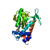
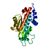
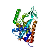
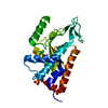
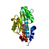
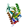
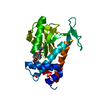
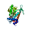
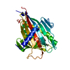
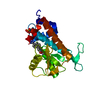
 RNA BINDING PROTEIN / CAP-SNATCHING
 Function and homology information

 Authors
 Citation
 Structure visualization
 Downloads & links
 Other downloads
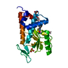


 PDBj
PDBj Assembly
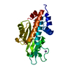


 Mass: 54.938 Da / Num. of mol.: 3 / Source method: obtained synthetically / Formula: Mn
 Mass: 62.068 Da / Num. of mol.: 1 / Source method: obtained synthetically / Formula: C2H6O2
 Mass: 18.015 Da / Num. of mol.: 225 / Source method: isolated from a natural source / Formula: H2O
 Sample preparation
 / Beamline: X25 / Wavelength: 1.1 / Wavelength: 1.1 Å
 Processing