 Movie
Movie Controller

[English] 日本語
 Yorodumi
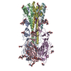
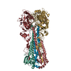
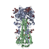
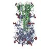
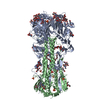
- PDB-4m40: Crystal structure of hemagglutinin of influenza virus B/Yamanashi... -
+ Open data

- Basic information
| Entry | Database: PDB / ID: 4m40 | |||||||||
|---|---|---|---|---|---|---|---|---|---|---|
| Title | Crystal structure of hemagglutinin of influenza virus B/Yamanashi/166/1998 | |||||||||
 Components Components |
| |||||||||
 Keywords Keywords |  VIRAL PROTEIN / receptor binding / fusion / sialic acid VIRAL PROTEIN / receptor binding / fusion / sialic acid | |||||||||
| Function / homology |  Function and homology informationviral budding from plasma membrane / endocytosis involved in viral entry into host cell / host cell surface receptor binding / apical plasma membrane / fusion of virus membrane with host plasma membrane / fusion of virus membrane with host endosome membrane / viral envelope / virion attachment to host cell / host cell plasma membrane / virion membrane Function and homology informationviral budding from plasma membrane / endocytosis involved in viral entry into host cell / host cell surface receptor binding / apical plasma membrane / fusion of virus membrane with host plasma membrane / fusion of virus membrane with host endosome membrane / viral envelope / virion attachment to host cell / host cell plasma membrane / virion membraneSimilarity search - Function | |||||||||
| Biological species |  Influenza B virus Influenza B virus | |||||||||
| Method | X-RAY DIFFRACTION / SYNCHROTRON / MOLECULAR REPLACEMENT / Resolution: 3.54 Å | |||||||||
 Authors Authors | Ni, F. / Kondrashkina, E. / Wang, Q. | |||||||||
 Citation Citation | Journal: Virology / Year: 2013 Title: Structural basis for the divergent evolution of influenza B virus hemagglutinin. Authors: Ni, F. / Kondrashkina, E. / Wang, Q. | |||||||||
| History |
|
- Structure visualization
| Structure viewer | Molecule: MolmilJmol/JSmol |
|---|
- Downloads & links
-Download
| PDBx/mmCIF format | 4m40.cif.gz | 300.2 KB | Display | PDBx/mmCIF format |
|---|---|---|---|---|
| PDB format | pdb4m40.ent.gz | 250.3 KB | Display | PDB format |
| PDBx/mmJSON format | 4m40.json.gz | Tree view | PDBx/mmJSON format | |
| Others |  Other downloads Other downloads |
-Validation report
| Arichive directory | https://data.pdbj.org/pub/pdb/validation_reports/m4/4m40ftp://data.pdbj.org/pub/pdb/validation_reports/m4/4m40 | HTTPS FTP |
|---|
-Related structure data


-Links
 PDBj
PDBj


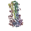
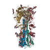
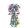
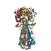
- Assembly
| Deposited unit | 
| ||||||||
|---|---|---|---|---|---|---|---|---|---|
| 1 |
| ||||||||
| Unit cell |
|
-Components
| #1: Protein | Mass: 37543.043 Da / Num. of mol.: 3 / Fragment: Hemagglutinin HA1 Source method: isolated from a genetically manipulated source Source: (gene. exp.) Influenza B virus / Strain: B/Yamanashi/166/1998 / Gene: HA / Plasmid: pRB21 / Production host: Mammalia (mammals) / Strain (production host): CV-1 / References: UniProt: A3DQM7#2: Protein | Mass: 19542.980 Da / Num. of mol.: 3 / Fragment: Hemagglutinin HA2 Source method: isolated from a genetically manipulated source Source: (gene. exp.) Influenza B virus / Strain: B/Yamanashi/166/1998 / Gene: HA / Plasmid: pRB21 / Production host: Mammalia (mammals) / Strain (production host): CV-1 / References: UniProt: A3DQM7#3: Polysaccharide | 2-acetamido-2-deoxy-beta-D-glucopyranose-(1-4)-2-acetamido-2-deoxy-beta-D-glucopyranose / Mass: 424.401 Da / Num. of mol.: 9Source method: isolated from a genetically manipulated source #4: Sugar | ChemComp-NAG / N-Acetylglucosamine  Type: D-saccharide, beta linking / Mass: 221.208 Da / Num. of mol.: 12 Type: D-saccharide, beta linking / Mass: 221.208 Da / Num. of mol.: 12Source method: isolated from a genetically manipulated source Formula: C8H15NO6 |
|---|
-Experimental details
-Experiment
| Experiment | Method: X-RAY DIFFRACTION / Number of used crystals: 1 |
|---|
- Sample preparation
| Crystal | Density Matthews: 3.2 Å3/Da / Density % sol: 61.59 % |
|---|---|
| Crystal grow | Temperature: 290 K / Method: vapor diffusion, hanging drop / pH: 8.5 Details: 23% PEG2000MME, 0.1 M Tris, 0.2 M TMNO, pH 8.5, VAPOR DIFFUSION, HANGING DROP, temperature 290K |
-Data collection
| Diffraction | Mean temperature: 70 K |
|---|---|
| Diffraction source | Source: SYNCHROTRON / Site: APS  / Beamline: 21-ID-G / Wavelength: 0.97857 Å / Beamline: 21-ID-G / Wavelength: 0.97857 Å |
| Detector | Type: RAYONIX MX-225 / Detector: CCD / Date: Jun 6, 2012 |
| Radiation | Protocol: SINGLE WAVELENGTH / Monochromatic (M) / Laue (L): M / Scattering type: x-ray |
| Radiation wavelength | Wavelength: 0.97857 Å / Relative weight: 1 |
| Reflection | Resolution: 3.54→43.6 Å / Num. all: 26547 / Num. obs: 26547 / % possible obs: 100 % / Observed criterion σ(F): 2 / Observed criterion σ(I): 1 / Redundancy: 3.8 % / Biso Wilson estimate: 109.1 Å2 / Rmerge(I) obs: 0.098 |
| Reflection shell | Resolution: 3.54→3.73 Å / Redundancy: 3.8 % / Rmerge(I) obs: 0.497 / Mean I/σ(I) obs: 2.3 / % possible all: 100 |
- Processing
| Software |
| |||||||||||||||||||||||||||||||||||||||||||||||||||||||||||||||||||||||||||||
|---|---|---|---|---|---|---|---|---|---|---|---|---|---|---|---|---|---|---|---|---|---|---|---|---|---|---|---|---|---|---|---|---|---|---|---|---|---|---|---|---|---|---|---|---|---|---|---|---|---|---|---|---|---|---|---|---|---|---|---|---|---|---|---|---|---|---|---|---|---|---|---|---|---|---|---|---|---|---|
| Refinement | Method to determine structure: MOLECULAR REPLACEMENT / Resolution: 3.54→42.66 Å / SU ML: 0.45 / σ(F): 1.49 / Phase error: 25.82 / Stereochemistry target values: ML
| |||||||||||||||||||||||||||||||||||||||||||||||||||||||||||||||||||||||||||||
| Solvent computation | Shrinkage radii: 0.9 Å / VDW probe radii: 1.11 Å / Solvent model: FLAT BULK SOLVENT MODEL | |||||||||||||||||||||||||||||||||||||||||||||||||||||||||||||||||||||||||||||
| Refinement step | Cycle: LAST / Resolution: 3.54→42.66 Å
| |||||||||||||||||||||||||||||||||||||||||||||||||||||||||||||||||||||||||||||
| Refine LS restraints |
| |||||||||||||||||||||||||||||||||||||||||||||||||||||||||||||||||||||||||||||
| LS refinement shell |
|
