[English] 日本語
Yorodumi
- PDB-4m3p: Betaine-Homocysteine S-Methyltransferase from Homo sapiens comple... -

+
Open data


ID or keywords:

Loading...

-
Basic information

Entry
Database: PDB / ID: 4m3p
TitleBetaine-Homocysteine S-Methyltransferase from Homo sapiens complexed with Homocysteine
ComponentsBetaine--homocysteine S-methyltransferase 1
KeywordsTRANSFERASE / Alpha and beta proteins (a/b) / TIM beta/alpha-barrel / methyltransferase activity / homocysteine S-methyltransferase activity / transferase activity / metal ion binding / betaine-homocysteine S-methyltransferase activity / protein complex binding / betaine / homocysteine
Function / homology
Function and homology information


regulation of homocysteine metabolic process / betaine-homocysteine S-methyltransferase / amino-acid betaine metabolic process / betaine-homocysteine S-methyltransferase activity / L-methionine salvage / amino-acid betaine catabolic process / Sulfur amino acid metabolism / Choline catabolism / 'de novo' L-methionine biosynthetic process / protein methylation ...regulation of homocysteine metabolic process / betaine-homocysteine S-methyltransferase / amino-acid betaine metabolic process / betaine-homocysteine S-methyltransferase activity / L-methionine salvage / amino-acid betaine catabolic process / Sulfur amino acid metabolism / Choline catabolism / 'de novo' L-methionine biosynthetic process / protein methylation / extracellular exosome / zinc ion binding / nucleus / cytosol
Similarity search - Function
Betaine-homocysteine S-methyltransferase, BHMT / Homocysteine-binding-like domain / Homocysteine-binding domain / Homocysteine-binding domain superfamily / Homocysteine S-methyltransferase / Homocysteine-binding domain profile. / TIM Barrel / Alpha-Beta Barrel / Alpha Beta
Similarity search - Domain/homology
2-AMINO-4-MERCAPTO-BUTYRIC ACID / : / S,R MESO-TARTARIC ACID / Betaine--homocysteine S-methyltransferase 1
Similarity search - Component
Biological speciesHomo sapiens (human)
MethodX-RAY DIFFRACTION / SYNCHROTRON / MOLECULAR REPLACEMENT / Resolution: 1.895 Å
AuthorsKoutmos, M. / Yamada, K. / Mladkova, J. / Paterova, J. / Diamond, C.E. / Tryon, K. / Jungwirth, P. / Garrow, T.A. / Jiracek, J.
CitationJournal: Proteins / Year: 2014
Title: Specific potassium ion interactions facilitate homocysteine binding to betaine-homocysteine S-methyltransferase.
Authors: Mladkova, J. / Hladilkova, J. / Diamond, C.E. / Tryon, K. / Yamada, K. / Garrow, T.A. / Jungwirth, P. / Koutmos, M. / Jiracek, J.
History
DepositionAug 6, 2013Deposition site: RCSB / Processing site: RCSB
Revision 1.0Jun 18, 2014Provider: repository / Type: Initial release
Revision 1.1Oct 8, 2014Group: Database references
Revision 1.2Sep 20, 2023Group: Data collection / Database references ...Data collection / Database references / Derived calculations / Refinement description
Category: chem_comp_atom / chem_comp_bond ...chem_comp_atom / chem_comp_bond / database_2 / pdbx_initial_refinement_model / pdbx_struct_conn_angle / struct_conn / struct_site
Item: _database_2.pdbx_DOI / _database_2.pdbx_database_accession ..._database_2.pdbx_DOI / _database_2.pdbx_database_accession / _pdbx_struct_conn_angle.ptnr1_auth_asym_id / _pdbx_struct_conn_angle.ptnr1_auth_comp_id / _pdbx_struct_conn_angle.ptnr1_auth_seq_id / _pdbx_struct_conn_angle.ptnr1_label_asym_id / _pdbx_struct_conn_angle.ptnr1_label_atom_id / _pdbx_struct_conn_angle.ptnr1_label_comp_id / _pdbx_struct_conn_angle.ptnr1_label_seq_id / _pdbx_struct_conn_angle.ptnr2_auth_asym_id / _pdbx_struct_conn_angle.ptnr2_auth_comp_id / _pdbx_struct_conn_angle.ptnr2_auth_seq_id / _pdbx_struct_conn_angle.ptnr2_label_asym_id / _pdbx_struct_conn_angle.ptnr2_label_atom_id / _pdbx_struct_conn_angle.ptnr2_label_comp_id / _pdbx_struct_conn_angle.ptnr3_auth_asym_id / _pdbx_struct_conn_angle.ptnr3_auth_comp_id / _pdbx_struct_conn_angle.ptnr3_auth_seq_id / _pdbx_struct_conn_angle.ptnr3_label_asym_id / _pdbx_struct_conn_angle.ptnr3_label_atom_id / _pdbx_struct_conn_angle.ptnr3_label_comp_id / _pdbx_struct_conn_angle.ptnr3_label_seq_id / _pdbx_struct_conn_angle.value / _struct_conn.pdbx_dist_value / _struct_conn.ptnr1_auth_asym_id / _struct_conn.ptnr1_auth_comp_id / _struct_conn.ptnr1_auth_seq_id / _struct_conn.ptnr1_label_asym_id / _struct_conn.ptnr1_label_atom_id / _struct_conn.ptnr1_label_comp_id / _struct_conn.ptnr1_label_seq_id / _struct_conn.ptnr2_auth_asym_id / _struct_conn.ptnr2_auth_comp_id / _struct_conn.ptnr2_auth_seq_id / _struct_conn.ptnr2_label_asym_id / _struct_conn.ptnr2_label_atom_id / _struct_conn.ptnr2_label_comp_id / _struct_site.pdbx_auth_asym_id / _struct_site.pdbx_auth_comp_id / _struct_site.pdbx_auth_seq_id
Revision 2.0Nov 15, 2023Group: Atomic model / Data collection / Category: atom_site / chem_comp_atom / chem_comp_bond
Item: _atom_site.auth_atom_id / _atom_site.label_atom_id ..._atom_site.auth_atom_id / _atom_site.label_atom_id / _chem_comp_atom.atom_id / _chem_comp_bond.atom_id_2
Revision 2.1Apr 3, 2024Group: Structure summary / Category: chem_comp / Item: _chem_comp.pdbx_synonyms

-
Structure visualization

Structure viewerMolecule:
MolmilJmol/JSmol

Downloads & links

-
Assembly

Deposited unit
A: Betaine--homocysteine S-methyltransferase 1
B: Betaine--homocysteine S-methyltransferase 1
C: Betaine--homocysteine S-methyltransferase 1
D: Betaine--homocysteine S-methyltransferase 1
hetero molecules


Theoretical massNumber of molelcules
Total (without water)181,38018
Polymers180,1224
Non-polymers1,25914
Water11,602644
1


  • Idetical with deposited unit
  • defined by author&software
TypeNameSymmetry operationNumber
identity operation1_555x,y,z1
Buried area20430 Å2
ΔGint-76 kcal/mol
Surface area54010 Å2
MethodPISA
Unit cell
Length a, b, c (Å)86.097, 102.617, 96.228
Angle α, β, γ (deg.)90.00, 101.76, 90.00
Int Tables number4
Space group name H-MP1211

-
Components

-
Protein , 1 types, 4 molecules ABCD

#1: Protein
Betaine--homocysteine S-methyltransferase 1


Mass: 45030.379 Da / Num. of mol.: 4
Source method: isolated from a genetically manipulated source
Source: (gene. exp.) Homo sapiens (human) / Gene: BHMT / Plasmid: pTYB4 / Production host: Escherichia coli (E. coli) / Strain (production host): BL21(DE3)
References: UniProt: Q93088, betaine-homocysteine S-methyltransferase

-
Non-polymers , 5 types, 658 molecules

#2: Chemical
ChemComp-ZN / ZINC ION


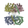
Mass: 65.409 Da / Num. of mol.: 4 / Source method: obtained synthetically / Formula: Zn
#3: Chemical
ChemComp-K / POTASSIUM ION


Mass: 39.098 Da / Num. of mol.: 4 / Source method: obtained synthetically / Formula: K
#4: Chemical
ChemComp-HCS / 2-AMINO-4-MERCAPTO-BUTYRIC ACID / L-Homocysteine / Homocysteine


Type: L-peptide linking / Mass: 135.185 Da / Num. of mol.: 4 / Source method: obtained synthetically / Formula: C4H9NO2S
#5: Chemical ChemComp-SRT / S,R MESO-TARTARIC ACID


Mass: 150.087 Da / Num. of mol.: 2 / Source method: obtained synthetically / Formula: C4H6O6
#6: Water ChemComp-HOH / water / Water


Mass: 18.015 Da / Num. of mol.: 644 / Source method: isolated from a natural source / Formula: H2O

-
Details

Sequence detailsR239Q IS A NATURAL VARIANT.

-
Experimental details

-
Experiment

ExperimentMethod: X-RAY DIFFRACTION / Number of used crystals: 1

-
Sample preparation

CrystalDensity Matthews: 2.31 Å3/Da / Density % sol: 46.76 %
Crystal growTemperature: 277 K / Method: evaporation / pH: 7.5
Details: 20.0% PEG5000 MME, 0.2 M sodium/potassium tartrate, 1.4% 1,2,3-heptanetriol, pH 7.5, EVAPORATION, temperature 277K

-
Data collection

DiffractionMean temperature: 100 K
Diffraction sourceSource: SYNCHROTRON / Site: APS / Beamline: 23-ID-D / Wavelength: 1.0332 Å
DetectorType: MARMOSAIC 300 mm CCD / Detector: CCD / Date: Dec 18, 2006
Details: K-B pair of biomorph mirrors for vertical and horizontal focusing
RadiationMonochromator: double crystal Si(111) / Protocol: SINGLE WAVELENGTH / Monochromatic (M) / Laue (L): M / Scattering type: x-ray
Radiation wavelengthWavelength: 1.0332 Å / Relative weight: 1
ReflectionResolution: 1.895→45.222 Å / Num. all: 129862 / Num. obs: 128582 / % possible obs: 98.2 % / Observed criterion σ(F): 0 / Observed criterion σ(I): 0 / Redundancy: 4.1 % / Biso Wilson estimate: 34.8 Å2 / Rmerge(I) obs: 0.083 / Rsym value: 0.072 / Net I/σ(I): 13.7
Reflection shellResolution: 1.895→2.01 Å / Redundancy: 4.1 % / Rmerge(I) obs: 0.374 / Mean I/σ(I) obs: 4 / Num. unique all: 19787 / Rsym value: 0.369 / % possible all: 90

-
Processing

Software
NameVersionClassification
Blu-Icedata collection
EPMRphasing
REFMAC5.6.0117refinement
XDSdata reduction
XSCALEdata scaling
RefinementMethod to determine structure: MOLECULAR REPLACEMENT
Starting model: PDB ENTRY 1LT8
Resolution: 1.895→45.222 Å / Cor.coef. Fo:Fc: 0.962 / Cor.coef. Fo:Fc free: 0.946 / SU B: 3.334 / SU ML: 0.097 / Cross valid method: THROUGHOUT / σ(F): 0 / σ(I): 0 / ESU R: 0.142 / ESU R Free: 0.134 / Stereochemistry target values: MAXIMUM LIKELIHOOD / Details: HYDROGENS HAVE BEEN USED IF PRESENT IN THE INPUT
RfactorNum. reflection% reflectionSelection details
Rfree0.21852 6476 5 %RANDOM
Rwork0.18021 ---
obs0.18214 122106 99.01 %-
all-123327 --
Solvent computationIon probe radii: 0.8 Å / Shrinkage radii: 0.8 Å / VDW probe radii: 1.2 Å / Solvent model: BABINET MODEL WITH MASK
Displacement parametersBiso mean: 33.943 Å2
Baniso -1Baniso -2Baniso -3
1--0.14 Å20 Å20.42 Å2
2--2.05 Å20 Å2
3----1.74 Å2
Refinement stepCycle: LAST / Resolution: 1.895→45.222 Å
ProteinNucleic acidLigandSolventTotal
Num. atoms11469 0 60 644 12173
Refine LS restraints
Refine-IDTypeDev idealDev ideal targetNumber
X-RAY DIFFRACTIONr_bond_refined_d0.0110.01911821
X-RAY DIFFRACTIONr_bond_other_d0.0010.0211346
X-RAY DIFFRACTIONr_angle_refined_deg1.3561.95915983
X-RAY DIFFRACTIONr_angle_other_deg0.806325980
X-RAY DIFFRACTIONr_dihedral_angle_1_deg5.51651463
X-RAY DIFFRACTIONr_dihedral_angle_2_deg32.21824.063539
X-RAY DIFFRACTIONr_dihedral_angle_3_deg15.009151989
X-RAY DIFFRACTIONr_dihedral_angle_4_deg17.9511572
X-RAY DIFFRACTIONr_chiral_restr0.0820.21687
X-RAY DIFFRACTIONr_gen_planes_refined0.0070.02113392
X-RAY DIFFRACTIONr_gen_planes_other0.0010.022636
LS refinement shellResolution: 1.895→1.944 Å / Total num. of bins used: 20
RfactorNum. reflection% reflection
Rfree0.274 395 -
Rwork0.246 7870 -
obs--87.71 %

+
About Yorodumi

-
News

-
Feb 9, 2022. New format data for meta-information of EMDB entries

New format data for meta-information of EMDB entries

  • Version 3 of the EMDB header file is now the official format.
  • The previous official version 1.9 will be removed from the archive.

Related info.:EMDB header

External links:wwPDB to switch to version 3 of the EMDB data model

-
Aug 12, 2020. Covid-19 info

Covid-19 info

URL: https://pdbj.org/emnavi/covid19.php

New page: Covid-19 featured information page in EM Navigator.

Related info.:Covid-19 info / Mar 5, 2020. Novel coronavirus structure data

+
Mar 5, 2020. Novel coronavirus structure data

Novel coronavirus structure data

Related info.:Yorodumi Speices / Aug 12, 2020. Covid-19 info

External links:COVID-19 featured content - PDBj / Molecule of the Month (242):Coronavirus Proteases

+
Jan 31, 2019. EMDB accession codes are about to change! (news from PDBe EMDB page)

EMDB accession codes are about to change! (news from PDBe EMDB page)

  • The allocation of 4 digits for EMDB accession codes will soon come to an end. Whilst these codes will remain in use, new EMDB accession codes will include an additional digit and will expand incrementally as the available range of codes is exhausted. The current 4-digit format prefixed with “EMD-” (i.e. EMD-XXXX) will advance to a 5-digit format (i.e. EMD-XXXXX), and so on. It is currently estimated that the 4-digit codes will be depleted around Spring 2019, at which point the 5-digit format will come into force.
  • The EM Navigator/Yorodumi systems omit the EMD- prefix.

Related info.:Q: What is EMD? / ID/Accession-code notation in Yorodumi/EM Navigator

External links:EMDB Accession Codes are Changing Soon! / Contact to PDBj

+
Jul 12, 2017. Major update of PDB

Major update of PDB

  • wwPDB released updated PDB data conforming to the new PDBx/mmCIF dictionary.
  • This is a major update changing the version number from 4 to 5, and with Remediation, in which all the entries are updated.
  • In this update, many items about electron microscopy experimental information are reorganized (e.g. em_software).
  • Now, EM Navigator and Yorodumi are based on the updated data.

External links:wwPDB Remediation / Enriched Model Files Conforming to OneDep Data Standards Now Available in the PDB FTP Archive

-
Yorodumi

Thousand views of thousand structures

  • Yorodumi is a browser for structure data from EMDB, PDB, SASBDB, etc.
  • This page is also the successor to EM Navigator detail page, and also detail information page/front-end page for Omokage search.
  • The word "yorodu" (or yorozu) is an old Japanese word meaning "ten thousand". "mi" (miru) is to see.

Related info.:EMDB / PDB / SASBDB / Comparison of 3 databanks / Yorodumi Search / Aug 31, 2016. New EM Navigator & Yorodumi / Yorodumi Papers / Jmol/JSmol / Function and homology information / Changes in new EM Navigator and Yorodumi

Read more