 Movie
Movie Controller

[English] 日本語
 Yorodumi
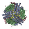
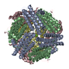
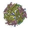
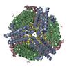
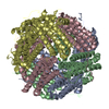
- PDB-4m35: Crystal structure of gated-pore mutant H126/141D of second DNA-Bi... -
+ Open data

- Basic information
| Entry | Database: PDB / ID: 4m35 | ||||||
|---|---|---|---|---|---|---|---|
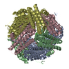
| Title | Crystal structure of gated-pore mutant H126/141D of second DNA-Binding protein under starvation from Mycobacterium smegmatis | ||||||
 Components Components | Putative starvation-induced DNA protecting protein/Ferritin and Dps | ||||||
 Keywords Keywords |  DNA BINDING PROTEIN / ferritin-like fold / DNA binding / ferroxidation / Iron DNA BINDING PROTEIN / ferritin-like fold / DNA binding / ferroxidation / Iron | ||||||
| Function / homology |  Function and homology information Function and homology informationoxidoreductase activity, acting on metal ions / ferric iron binding / DNA bindingSimilarity search - Function | ||||||
| Biological species |  Mycobacterium smegmatis (bacteria) Mycobacterium smegmatis (bacteria) | ||||||
| Method | X-RAY DIFFRACTION / Direct refinement against model PDB / Resolution: 2.05 Å | ||||||
 Authors Authors | Williams, S.M. / Chandran, A.V. / Vijayabaskar, M.S. / Roy, S. / Balaram, H. / Vishveshwara, S. / Vijayan, M. / Chatterji, D. | ||||||
 Citation Citation | Journal: J.Biol.Chem. / Year: 2014 Title: A histidine aspartate ionic lock gates the iron passage in miniferritins from Mycobacterium smegmatis Authors: Williams, S.M. / Chandran, A.V. / Vijayabaskar, M.S. / Roy, S. / Balaram, H. / Vishveshwara, S. / Vijayan, M. / Chatterji, D. | ||||||
| History |
|
- Structure visualization
| Structure viewer | Molecule: MolmilJmol/JSmol |
|---|
- Downloads & links
-Download
| PDBx/mmCIF format | 4m35.cif.gz | 139.1 KB | Display | PDBx/mmCIF format |
|---|---|---|---|---|
| PDB format | pdb4m35.ent.gz | 108.9 KB | Display | PDB format |
| PDBx/mmJSON format | 4m35.json.gz | Tree view | PDBx/mmJSON format | |
| Others |  Other downloads Other downloads |
-Validation report
| Arichive directory | https://data.pdbj.org/pub/pdb/validation_reports/m3/4m35ftp://data.pdbj.org/pub/pdb/validation_reports/m3/4m35 | HTTPS FTP |
|---|
-Related structure data
| Related structure data | 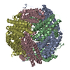 4m32C 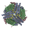 4m33C 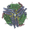 4m34C  2z90S C: citing same article ( S: Starting model for refinement |
|---|---|
| Similar structure data |


-Links
 PDBj
PDBj


- Assembly
| Deposited unit | 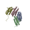
| |||||||||||||||
|---|---|---|---|---|---|---|---|---|---|---|---|---|---|---|---|---|
| 1 | 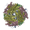
| |||||||||||||||
| Unit cell |
| |||||||||||||||
| Components on special symmetry positions |
|
-Components
| #1: Protein | Mass: 18466.568 Da / Num. of mol.: 4 / Mutation: H126D, H141D Source method: isolated from a genetically manipulated source Source: (gene. exp.) Mycobacterium smegmatis (bacteria) / Strain: ATCC 700084 / mc(2)155 / Gene: DPS2, MSMEG_3242, MSMEI_3159 / Plasmid: pET21b / Production host: Escherichia coli (E. coli) / Strain (production host): BL21(DE3) / References: UniProt: A0QXB7#2: Chemical | ChemComp-FE2 /   Mass: 55.845 Da / Num. of mol.: 4 / Source method: obtained synthetically / Formula: Fe Mass: 55.845 Da / Num. of mol.: 4 / Source method: obtained synthetically / Formula: Fe#3: Chemical |   Mass: 24.305 Da / Num. of mol.: 2 / Source method: obtained synthetically / Formula: Mg Mass: 24.305 Da / Num. of mol.: 2 / Source method: obtained synthetically / Formula: Mg#4: Chemical | Chloride  Mass: 35.453 Da / Num. of mol.: 2 / Source method: obtained synthetically / Formula: Cl Mass: 35.453 Da / Num. of mol.: 2 / Source method: obtained synthetically / Formula: Cl#5: Water | ChemComp-HOH / | Water Mass: 18.015 Da / Num. of mol.: 384 / Source method: isolated from a natural source / Formula: H2O Mass: 18.015 Da / Num. of mol.: 384 / Source method: isolated from a natural source / Formula: H2O |
|---|
-Experimental details
-Experiment
| Experiment | Method: X-RAY DIFFRACTION / Number of used crystals: 1 |
|---|
- Sample preparation
| Crystal | Density Matthews: 2.23 Å3/Da / Density % sol: 44.81 % Description: THE ENTRY CONTAINS FRIEDEL PAIRS IN F_PLUS/MINUS COLUMNS. |
|---|---|
| Crystal grow | Temperature: 298 K / Method: microbatch under oil / pH: 6.5 Details: 50mM MgCl2, 0.1M sodium cacodylate, 20% PEG3350 , pH 6.5, Microbatch under oil, temperature 298K |
-Data collection
| Diffraction | Mean temperature: 100 K |
|---|---|
| Diffraction source | Source: ROTATING ANODE / Type: BRUKER AXS MICROSTAR / Wavelength: 1.54179 Å |
| Detector | Type: MAR scanner 345 mm plate / Detector: IMAGE PLATE / Date: Jul 22, 2011 |
| Radiation | Monochromator: Osmic / Protocol: SINGLE WAVELENGTH / Monochromatic (M) / Laue (L): M / Scattering type: x-ray |
| Radiation wavelength | Wavelength: 1.54179 Å / Relative weight: 1 |
| Reflection | Resolution: 2.03→30 Å / Num. obs: 41264 / Redundancy: 7 % / Biso Wilson estimate: 13.1 Å2 / Rmerge(I) obs: 0.053 / Net I/σ(I): 25.6 |
| Reflection shell | Resolution: 2.03→2.14 Å / Redundancy: 6.4 % / Rmerge(I) obs: 0.139 / Mean I/σ(I) obs: 11.7 |
- Processing
| Software |
| |||||||||||||||||||||||||||||||||||||||||||||||||||||||||||||||||
|---|---|---|---|---|---|---|---|---|---|---|---|---|---|---|---|---|---|---|---|---|---|---|---|---|---|---|---|---|---|---|---|---|---|---|---|---|---|---|---|---|---|---|---|---|---|---|---|---|---|---|---|---|---|---|---|---|---|---|---|---|---|---|---|---|---|---|
| Refinement | Method to determine structure: Direct refinement against model PDB Starting model: 2z90 Resolution: 2.05→30 Å / Cor.coef. Fo:Fc: 0.941 / Cor.coef. Fo:Fc free: 0.92 / SU B: 3.424 / SU ML: 0.095 / Cross valid method: THROUGHOUT / ESU R: 0.2 / ESU R Free: 0.16 / Stereochemistry target values: MAXIMUM LIKELIHOOD Details: SF FILE CONTAINS FRIEDEL PAIRS UNDER I/F_MINUS AND I/F_PLUS COLUMNS.
| |||||||||||||||||||||||||||||||||||||||||||||||||||||||||||||||||
| Solvent computation | Ion probe radii: 0.8 Å / Shrinkage radii: 0.8 Å / VDW probe radii: 1.4 Å / Solvent model: MASK | |||||||||||||||||||||||||||||||||||||||||||||||||||||||||||||||||
| Displacement parameters | Biso mean: 12.356 Å2
| |||||||||||||||||||||||||||||||||||||||||||||||||||||||||||||||||
| Refinement step | Cycle: LAST / Resolution: 2.05→30 Å
| |||||||||||||||||||||||||||||||||||||||||||||||||||||||||||||||||
| Refine LS restraints |
| |||||||||||||||||||||||||||||||||||||||||||||||||||||||||||||||||
| LS refinement shell | Resolution: 2.05→2.106 Å / Total num. of bins used: 20
|
