 Movie
Movie Controller

[English] 日本語
 Yorodumi
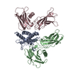
- PDB-4m0m: The crystal structure of a functionally unknown protein lpg2422 f... -
+ Open data

- Basic information
| Entry | Database: PDB / ID: 4m0m | ||||||
|---|---|---|---|---|---|---|---|
| Title | The crystal structure of a functionally unknown protein lpg2422 from Legionella pneumophila subsp. pneumophila str. Philadelphia 1 | ||||||
 Components Components | Putative uncharacterized protein | ||||||
 Keywords Keywords | UNKNOWN FUNCTION /  structural genomics / PSI-Biology / protein structure initiative / Midwest Center for Structural Genomics / MCSG structural genomics / PSI-Biology / protein structure initiative / Midwest Center for Structural Genomics / MCSG | ||||||
| Function / homology |  Function and homology informationMethyltransferase, Methionine Synthase (B12-binding Domains); Chain A, domain 1 - #80 / Substrate Binding Domain Of Dnak; Chain:A; Domain 2 - #430 / Substrate Binding Domain Of Dnak; Chain:A; Domain 2 - #440 / Domain of unknown function DUF5621 / Domain of unknown function (DUF5621) / Methyltransferase, Methionine Synthase (B12-binding Domains); Chain A, domain 1 / Substrate Binding Domain Of Dnak; Chain:A; Domain 2 / Alpha/Beta hydrolase fold / Up-down Bundle / Orthogonal Bundle / Mainly Alpha Function and homology informationMethyltransferase, Methionine Synthase (B12-binding Domains); Chain A, domain 1 - #80 / Substrate Binding Domain Of Dnak; Chain:A; Domain 2 - #430 / Substrate Binding Domain Of Dnak; Chain:A; Domain 2 - #440 / Domain of unknown function DUF5621 / Domain of unknown function (DUF5621) / Methyltransferase, Methionine Synthase (B12-binding Domains); Chain A, domain 1 / Substrate Binding Domain Of Dnak; Chain:A; Domain 2 / Alpha/Beta hydrolase fold / Up-down Bundle / Orthogonal Bundle / Mainly AlphaSimilarity search - Domain/homology | ||||||
| Biological species |  Legionella pneumophila subsp. pneumophila (bacteria) Legionella pneumophila subsp. pneumophila (bacteria) | ||||||
| Method | X-RAY DIFFRACTION / SYNCHROTRON / SAD / Resolution: 2.192 Å | ||||||
 Authors Authors | Tan, K. / Li, H. / Clancy, S. / Shuman, H. / Joachimiak, A. / Midwest Center for Structural Genomics (MCSG) | ||||||
 Citation Citation | Journal: To be Published Title: The crystal structure of a functionally unknown protein lpg2422 from Legionella pneumophila subsp. pneumophila str. Philadelphia 1 Authors: Tan, K. / Li, H. / Clancy, S. / Shuman, H. / Joachimiak, A. | ||||||
| History |
|
- Structure visualization
| Structure viewer | Molecule: MolmilJmol/JSmol |
|---|
- Downloads & links
-Download
| PDBx/mmCIF format | 4m0m.cif.gz | 298.7 KB | Display | PDBx/mmCIF format |
|---|---|---|---|---|
| PDB format | pdb4m0m.ent.gz | 251.5 KB | Display | PDB format |
| PDBx/mmJSON format | 4m0m.json.gz | Tree view | PDBx/mmJSON format | |
| Others |  Other downloads Other downloads |
-Validation report
| Arichive directory | https://data.pdbj.org/pub/pdb/validation_reports/m0/4m0mftp://data.pdbj.org/pub/pdb/validation_reports/m0/4m0m | HTTPS FTP |
|---|
-Related structure data
| Similar structure data | |
|---|---|
| Other databases |

-Links
 PDBj
PDBj- Assembly
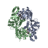
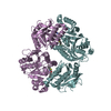
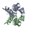
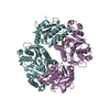
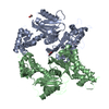
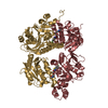
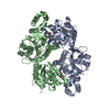
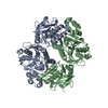
| Deposited unit | 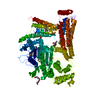
| ||||||||
|---|---|---|---|---|---|---|---|---|---|
| 1 |
| ||||||||
| Unit cell |
|
-Components
| #1: Protein | Mass: 87899.516 Da / Num. of mol.: 1 Source method: isolated from a genetically manipulated source Source: (gene. exp.) Legionella pneumophila subsp. pneumophila (bacteria)Strain: Philadelphia 1 / Gene: lpg2422 / Plasmid: pMCSG7 / Production host: Escherichia coli (E. coli) / Strain (production host): BL21(DE3)MAGIC / References: UniProt: Q5ZSU4 | ||||
|---|---|---|---|---|---|
| #2: Chemical | Polyethylene glycol  Mass: 194.226 Da / Num. of mol.: 3 / Source method: obtained synthetically / Formula: C8H18O5 / Comment: precipitant*YM Mass: 194.226 Da / Num. of mol.: 3 / Source method: obtained synthetically / Formula: C8H18O5 / Comment: precipitant*YM#3: Chemical | Phosphate  Mass: 94.971 Da / Num. of mol.: 2 / Source method: obtained synthetically / Formula: PO4 Mass: 94.971 Da / Num. of mol.: 2 / Source method: obtained synthetically / Formula: PO4#4: Water | ChemComp-HOH / | Water Mass: 18.015 Da / Num. of mol.: 82 / Source method: isolated from a natural source / Formula: H2O Mass: 18.015 Da / Num. of mol.: 82 / Source method: isolated from a natural source / Formula: H2O |
-Experimental details
-Experiment
| Experiment | Method: X-RAY DIFFRACTION / Number of used crystals: 1 |
|---|
- Sample preparation
| Crystal | Density Matthews: 2.39 Å3/Da / Density % sol: 48.58 % |
|---|---|
| Crystal grow | Temperature: 289 K / Method: vapor diffusion, sitting drop / pH: 6.5 Details: 0.2M Calcium Acetate, 0.1M Sodium Cacodylate:HCl, 40% PEG300, pH 6.5, VAPOR DIFFUSION, SITTING DROP, temperature 289K |
-Data collection
| Diffraction | Mean temperature: 100 K |
|---|---|
| Diffraction source | Source: SYNCHROTRON / Site: APS  / Beamline: 19-ID / Wavelength: 0.97937 Å / Beamline: 19-ID / Wavelength: 0.97937 Å |
| Detector | Type: ADSC QUANTUM 315r / Detector: CCD / Date: Aug 1, 2012 / Details: mirror |
| Radiation | Monochromator: Si 111 crystal / Protocol: SINGLE WAVELENGTH / Monochromatic (M) / Laue (L): M / Scattering type: x-ray |
| Radiation wavelength | Wavelength: 0.97937 Å / Relative weight: 1 |
| Reflection | Resolution: 2.2→34 Å / Num. all: 43629 / Num. obs: 43629 / % possible obs: 99.1 % / Observed criterion σ(F): -5 / Observed criterion σ(I): 0 / Redundancy: 4 % / Rmerge(I) obs: 0.095 / Net I/σ(I): 21.8 |
| Reflection shell | Resolution: 2.2→2.24 Å / Redundancy: 3.7 % / Rmerge(I) obs: 0.644 / Mean I/σ(I) obs: 1.6 / Num. unique all: 2037 / % possible all: 93.7 |
- Processing
| Software |
| |||||||||||||||||||||||||||||||||||||||||||||||||||||||||||||||||||||||||||||||||||||||||||||||||||||||||||||||||||||||||||||
|---|---|---|---|---|---|---|---|---|---|---|---|---|---|---|---|---|---|---|---|---|---|---|---|---|---|---|---|---|---|---|---|---|---|---|---|---|---|---|---|---|---|---|---|---|---|---|---|---|---|---|---|---|---|---|---|---|---|---|---|---|---|---|---|---|---|---|---|---|---|---|---|---|---|---|---|---|---|---|---|---|---|---|---|---|---|---|---|---|---|---|---|---|---|---|---|---|---|---|---|---|---|---|---|---|---|---|---|---|---|---|---|---|---|---|---|---|---|---|---|---|---|---|---|---|---|---|
| Refinement | Method to determine structure: SAD / Resolution: 2.192→33.975 Å / SU ML: 0.26 / σ(F): 1.33 / Phase error: 27.55 / Stereochemistry target values: ML
| |||||||||||||||||||||||||||||||||||||||||||||||||||||||||||||||||||||||||||||||||||||||||||||||||||||||||||||||||||||||||||||
| Solvent computation | Shrinkage radii: 0.9 Å / VDW probe radii: 1.11 Å / Solvent model: FLAT BULK SOLVENT MODEL | |||||||||||||||||||||||||||||||||||||||||||||||||||||||||||||||||||||||||||||||||||||||||||||||||||||||||||||||||||||||||||||
| Refinement step | Cycle: LAST / Resolution: 2.192→33.975 Å
| |||||||||||||||||||||||||||||||||||||||||||||||||||||||||||||||||||||||||||||||||||||||||||||||||||||||||||||||||||||||||||||
| Refine LS restraints |
| |||||||||||||||||||||||||||||||||||||||||||||||||||||||||||||||||||||||||||||||||||||||||||||||||||||||||||||||||||||||||||||
| LS refinement shell |
| |||||||||||||||||||||||||||||||||||||||||||||||||||||||||||||||||||||||||||||||||||||||||||||||||||||||||||||||||||||||||||||
| Refinement TLS params. | Method: refined / Refine-ID: X-RAY DIFFRACTION
| |||||||||||||||||||||||||||||||||||||||||||||||||||||||||||||||||||||||||||||||||||||||||||||||||||||||||||||||||||||||||||||
| Refinement TLS group |
|
