 Movie
Movie Controller


+ Open data

- Basic information
| Entry | Database: PDB / ID: 4lwd | ||||||
|---|---|---|---|---|---|---|---|
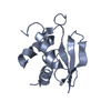
| Title | Human CARMA1 CARD domain | ||||||
 Components Components | Caspase recruitment domain-containing protein 11 | ||||||
 Keywords Keywords |  SIGNALING PROTEIN / death domain / Bcl10 SIGNALING PROTEIN / death domain / Bcl10 | ||||||
| Function / homology |  Function and homology information Function and homology informationthymic T cell selection / CBM complex / regulation of B cell differentiation / guanylate kinase activity / CD4-positive, alpha-beta T cell proliferation / TORC1 signaling / CARD domain binding / positive regulation of CD4-positive, alpha-beta T cell proliferation / regulation of T cell differentiation / positive regulation of T cell receptor signaling pathway ...thymic T cell selection / CBM complex / regulation of B cell differentiation / guanylate kinase activity / CD4-positive, alpha-beta T cell proliferation / TORC1 signaling / CARD domain binding / positive regulation of CD4-positive, alpha-beta T cell proliferation / regulation of T cell differentiation / positive regulation of T cell receptor signaling pathway / B cell proliferation / immunological synapse / homeostasis of number of cells / canonical NF-kappaB signal transduction / positive regulation of B cell proliferation / T cell costimulation / positive regulation of interleukin-2 production / B cell differentiation / Activation of NF-kappaB in B cells / protein homooligomerization / CLEC7A (Dectin-1) signaling / FCERI mediated NF-kB activation / : / Downstream TCR signaling / positive regulation of NF-kappaB transcription factor activity / regulation of apoptotic process / positive regulation of canonical NF-kappaB signal transduction / membrane raft / extracellular exosome / plasma membrane / cytosol / cytoplasmSimilarity search - Function | ||||||
| Biological species |  Homo sapiens (human) Homo sapiens (human) | ||||||
| Method | X-RAY DIFFRACTION / SAD / Resolution: 1.792 Å | ||||||
 Authors Authors | Zheng, C. / Wu, H. | ||||||
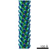
 Citation Citation | Journal: Mol Cell / Year: 2013 Title: Structural architecture of the CARMA1/Bcl10/MALT1 signalosome: nucleation-induced filamentous assembly. Authors: Qi Qiao / Chenghua Yang / Chao Zheng / Lorena Fontán / Liron David / Xiong Yu / Clay Bracken / Monica Rosen / Ari Melnick / Edward H Egelman / Hao Wu /  Abstract: The CARMA1/Bcl10/MALT1 (CBM) signalosome mediates antigen receptor-induced NF-κB signaling to regulate multiple lymphocyte functions. While CARMA1 and Bcl10 contain caspase recruitment domains ...The CARMA1/Bcl10/MALT1 (CBM) signalosome mediates antigen receptor-induced NF-κB signaling to regulate multiple lymphocyte functions. While CARMA1 and Bcl10 contain caspase recruitment domains (CARDs), MALT1 is a paracaspase with structural similarity to caspases. Here we show that the reconstituted CBM signalosome is a helical filamentous assembly in which substoichiometric CARMA1 nucleates Bcl10 filaments. Bcl10 filament formation is a highly cooperative process whose threshold is sensitized by oligomerized CARMA1 upon receptor activation. In cells, both cotransfected CARMA1/Bcl10 complex and the endogenous CBM signalosome are filamentous morphologically. Combining crystallography, nuclear magnetic resonance, and electron microscopy, we reveal the structure of the Bcl10 CARD filament and the mode of interaction between CARMA1 and Bcl10. Structure-guided mutagenesis confirmed the observed interfaces in Bcl10 filament assembly and MALT1 activation in vitro and NF-κB activation in cells. These data support a paradigm of nucleation-induced signal transduction with threshold response due to cooperativity and signal amplification by polymerization. | ||||||
| History |
|
- Structure visualization
| Structure viewer | Molecule: MolmilJmol/JSmol |
|---|
- Downloads & links
-Download
| PDBx/mmCIF format | 4lwd.cif.gz | 53 KB | Display | PDBx/mmCIF format |
|---|---|---|---|---|
| PDB format | pdb4lwd.ent.gz | 39.1 KB | Display | PDB format |
| PDBx/mmJSON format | 4lwd.json.gz | Tree view | PDBx/mmJSON format | |
| Others |  Other downloads Other downloads |
-Validation report
| Arichive directory | https://data.pdbj.org/pub/pdb/validation_reports/lw/4lwdftp://data.pdbj.org/pub/pdb/validation_reports/lw/4lwd | HTTPS FTP |
|---|
-Related structure data


-Links
 PDBj
PDBj


- Assembly
| Deposited unit | 
| ||||||||||||
|---|---|---|---|---|---|---|---|---|---|---|---|---|---|
| 1 |
| ||||||||||||
| 2 | x 6
| ||||||||||||
| Unit cell |
| ||||||||||||
| Components on special symmetry positions |
|
-Components
| #1: Protein | Mass: 11385.998 Da / Num. of mol.: 1 / Fragment: CARD domain residues 18-110 Source method: isolated from a genetically manipulated source Source: (gene. exp.) Homo sapiens (human) / Gene: CARD11, CARMA1 / Plasmid: pET28a / Production host:  Escherichia coli (E. coli) / Strain (production host): DE3 / References: UniProt: Q9BXL7 Escherichia coli (E. coli) / Strain (production host): DE3 / References: UniProt: Q9BXL7 | ||||
|---|---|---|---|---|---|
| #2: Chemical | Sulfate  Mass: 96.063 Da / Num. of mol.: 3 / Source method: obtained synthetically / Formula: SO4 Mass: 96.063 Da / Num. of mol.: 3 / Source method: obtained synthetically / Formula: SO4#3: Chemical | ChemComp-MG / |   Mass: 24.305 Da / Num. of mol.: 1 / Source method: obtained synthetically / Formula: Mg Mass: 24.305 Da / Num. of mol.: 1 / Source method: obtained synthetically / Formula: Mg#4: Water | ChemComp-HOH / | Water Mass: 18.015 Da / Num. of mol.: 60 / Source method: isolated from a natural source / Formula: H2O Mass: 18.015 Da / Num. of mol.: 60 / Source method: isolated from a natural source / Formula: H2O |
-Experimental details
-Experiment
| Experiment | Method: X-RAY DIFFRACTION / Number of used crystals: 1 |
|---|
- Sample preparation
| Crystal | Density Matthews: 2.02 Å3/Da / Density % sol: 39.14 % |
|---|---|
| Crystal grow | Temperature: 293 K / Method: vapor diffusion, hanging drop / pH: 6.5 Details: It was crystallized by mixing 1 to l protein (10 mg/ml in 20 mM Tris at pH 8.0, 150 mM NaCl, and 5 mM DTT) with 1 to l of the reservoir solution containing 1.4 M MgSO4 and 100 mM MES at pH 6. ...Details: It was crystallized by mixing 1 to l protein (10 mg/ml in 20 mM Tris at pH 8.0, 150 mM NaCl, and 5 mM DTT) with 1 to l of the reservoir solution containing 1.4 M MgSO4 and 100 mM MES at pH 6.5 in a hanging drop vapor diffusion system at 20 oC., VAPOR DIFFUSION, HANGING DROP, temperature 293K |
-Data collection
| Diffraction | Mean temperature: 100 K | ||||||||||||
|---|---|---|---|---|---|---|---|---|---|---|---|---|---|
| Diffraction source | Source: ROTATING ANODE / Type: RIGAKU / Wavelength: 1.5418 Å | ||||||||||||
| Detector | Type: RIGAKU RAXIS IV / Detector: IMAGE PLATE / Date: May 13, 2013 | ||||||||||||
| Radiation | Protocol: SINGLE WAVELENGTH / Monochromatic (M) / Laue (L): M / Scattering type: x-ray | ||||||||||||
| Radiation wavelength | Wavelength: 1.5418 Å / Relative weight: 1 | ||||||||||||
| Reflection | Resolution: 1.792→25.945 Å / Num. obs: 9328 / Observed criterion σ(F): 1.36 / Observed criterion σ(I): 2 | ||||||||||||
| Reflection shell |
|
- Processing
| Software |
| ||||||||||||||||||||||||||||||||||||||||||||||||||||||||||||||||||||||||||||||||||||||||||||||||||||||||||||||||||||||||||||||||||||||||||||||||||||||
|---|---|---|---|---|---|---|---|---|---|---|---|---|---|---|---|---|---|---|---|---|---|---|---|---|---|---|---|---|---|---|---|---|---|---|---|---|---|---|---|---|---|---|---|---|---|---|---|---|---|---|---|---|---|---|---|---|---|---|---|---|---|---|---|---|---|---|---|---|---|---|---|---|---|---|---|---|---|---|---|---|---|---|---|---|---|---|---|---|---|---|---|---|---|---|---|---|---|---|---|---|---|---|---|---|---|---|---|---|---|---|---|---|---|---|---|---|---|---|---|---|---|---|---|---|---|---|---|---|---|---|---|---|---|---|---|---|---|---|---|---|---|---|---|---|---|---|---|---|---|---|---|
| Refinement | Method to determine structure: SAD / Resolution: 1.792→25.945 Å / SU ML: 0.46 / σ(F): 1.36 / Phase error: 18.94 / Stereochemistry target values: ML
| ||||||||||||||||||||||||||||||||||||||||||||||||||||||||||||||||||||||||||||||||||||||||||||||||||||||||||||||||||||||||||||||||||||||||||||||||||||||
| Solvent computation | Shrinkage radii: 0.9 Å / VDW probe radii: 1.11 Å / Solvent model: FLAT BULK SOLVENT MODEL / Bsol: 55.146 Å2 / ksol: 0.449 e/Å3 | ||||||||||||||||||||||||||||||||||||||||||||||||||||||||||||||||||||||||||||||||||||||||||||||||||||||||||||||||||||||||||||||||||||||||||||||||||||||
| Displacement parameters |
| ||||||||||||||||||||||||||||||||||||||||||||||||||||||||||||||||||||||||||||||||||||||||||||||||||||||||||||||||||||||||||||||||||||||||||||||||||||||
| Refinement step | Cycle: LAST / Resolution: 1.792→25.945 Å
| ||||||||||||||||||||||||||||||||||||||||||||||||||||||||||||||||||||||||||||||||||||||||||||||||||||||||||||||||||||||||||||||||||||||||||||||||||||||
| Refine LS restraints |
| ||||||||||||||||||||||||||||||||||||||||||||||||||||||||||||||||||||||||||||||||||||||||||||||||||||||||||||||||||||||||||||||||||||||||||||||||||||||
| LS refinement shell |
| ||||||||||||||||||||||||||||||||||||||||||||||||||||||||||||||||||||||||||||||||||||||||||||||||||||||||||||||||||||||||||||||||||||||||||||||||||||||
| Refinement TLS params. | Method: refined / Refine-ID: X-RAY DIFFRACTION
| ||||||||||||||||||||||||||||||||||||||||||||||||||||||||||||||||||||||||||||||||||||||||||||||||||||||||||||||||||||||||||||||||||||||||||||||||||||||
| Refinement TLS group |
|
