 Movie
Movie Controller


+ Open data

- Basic information
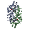
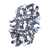
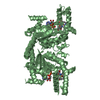
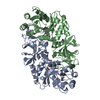
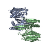
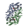
| Entry | Database: PDB / ID: 4lus | ||||||
|---|---|---|---|---|---|---|---|
| Title | alanine racemase [Clostridium difficile 630] | ||||||
 Components Components | (Alanine racemase ) x 2 ) x 2 | ||||||
 Keywords Keywords | ISOMERASE / Alanine racemase | ||||||
| Function / homology |  Function and homology informationalanine racemase / D-alanine biosynthetic process / alanine racemase activity / peptidoglycan biosynthetic process / pyridoxal phosphate binding / cytosol Function and homology informationalanine racemase / D-alanine biosynthetic process / alanine racemase activity / peptidoglycan biosynthetic process / pyridoxal phosphate binding / cytosolSimilarity search - Function | ||||||
| Biological species |  Clostridium difficile (bacteria) Clostridium difficile (bacteria) | ||||||
| Method | X-RAY DIFFRACTION / MOLECULAR REPLACEMENT / Resolution: 2.1 Å | ||||||
 Authors Authors | Asojo, O.A. | ||||||
 Citation Citation | Journal: Acta Crystallogr.,Sect.D / Year: 2014 Title: Structural and biochemical analyses of alanine racemase from the multidrug-resistant Clostridium difficile strain 630. Authors: Asojo, O.A. / Nelson, S.K. / Mootien, S. / Lee, Y. / Rezende, W.C. / Hyman, D.A. / Matsumoto, M.M. / Reiling, S. / Kelleher, A. / Ledizet, M. / Koski, R.A. / Anthony, K.G. | ||||||
| History |
|
- Structure visualization
| Structure viewer | Molecule: MolmilJmol/JSmol |
|---|
- Downloads & links
-Download
| PDBx/mmCIF format | 4lus.cif.gz | 314.9 KB | Display | PDBx/mmCIF format |
|---|---|---|---|---|
| PDB format | pdb4lus.ent.gz | 253.3 KB | Display | PDB format |
| PDBx/mmJSON format | 4lus.json.gz | Tree view | PDBx/mmJSON format | |
| Others |  Other downloads Other downloads |
-Validation report
| Arichive directory | https://data.pdbj.org/pub/pdb/validation_reports/lu/4lusftp://data.pdbj.org/pub/pdb/validation_reports/lu/4lus | HTTPS FTP |
|---|
-Related structure data
| Related structure data |  4lutC  4luyC  4a3qS S: Starting model for refinement C: citing same article ( |
|---|---|
| Similar structure data |


-Links
 PDBj
PDBj- Assembly
| Deposited unit | 
| ||||||||
|---|---|---|---|---|---|---|---|---|---|
| 1 | 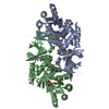
| ||||||||
| 2 | 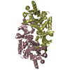
| ||||||||
| Unit cell |
|
-Components
| #1: Protein | Mass: 43636.066 Da / Num. of mol.: 3 Source method: isolated from a genetically manipulated source Details: Carboxylic modified Lysine / Source: (gene. exp.) Clostridium difficile (bacteria) / Strain: 630 / Gene: alr2, CD630_34630 / Production host:  Pichia pastoris (fungus) / References: UniProt: Q180W0, alanine racemase Pichia pastoris (fungus) / References: UniProt: Q180W0, alanine racemase#2: Protein | | Mass: 43593.066 Da / Num. of mol.: 1 Source method: isolated from a genetically manipulated source Source: (gene. exp.) Clostridium difficile (bacteria) / Strain: 630 / Gene: alr2, CD630_34630 / Production host: Pichia pastoris (fungus) / References: UniProt: Q180W0, alanine racemase#3: Chemical | ChemComp-GOL / | Glycerol  Mass: 92.094 Da / Num. of mol.: 1 / Source method: obtained synthetically / Formula: C3H8O3 Mass: 92.094 Da / Num. of mol.: 1 / Source method: obtained synthetically / Formula: C3H8O3#4: Chemical | ChemComp-TCE / | TCEP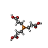  Mass: 250.186 Da / Num. of mol.: 1 / Source method: obtained synthetically / Formula: C9H15O6P Mass: 250.186 Da / Num. of mol.: 1 / Source method: obtained synthetically / Formula: C9H15O6P#5: Water | ChemComp-HOH / | Water Mass: 18.015 Da / Num. of mol.: 568 / Source method: isolated from a natural source / Formula: H2O Mass: 18.015 Da / Num. of mol.: 568 / Source method: isolated from a natural source / Formula: H2O |
|---|
-Experimental details
-Experiment
| Experiment | Method: X-RAY DIFFRACTION / Number of used crystals: 1 |
|---|
- Sample preparation
| Crystal | Density Matthews: 2.44 Å3/Da / Density % sol: 49.64 % |
|---|---|
| Crystal grow | Temperature: 298 K / pH: 7.5 Details: 20 mg/ml protein in 50mM Tris pH 8.0, 0.02% v/v BME, 10 micromolar pyridoxal-L-phosphate, crystallization buffer 1mM TCEP, 0.2M sodium formate, 20% w/V PEG3350, VAPOR DIFFUSION, SITTING DROP, temperature 298K |
-Data collection
| Diffraction | Mean temperature: 100 K |
|---|---|
| Diffraction source | Source: ROTATING ANODE / Type: RIGAKU FR-E+ SUPERBRIGHT / Wavelength: 1.54178 |
| Detector | Type: RIGAKU RAXIS HTC / Detector: IMAGE PLATE / Date: Sep 25, 2012 |
| Radiation | Protocol: SINGLE WAVELENGTH / Monochromatic (M) / Laue (L): M / Scattering type: x-ray |
| Radiation wavelength | Wavelength: 1.54178 Å / Relative weight: 1 |
| Reflection | Resolution: 2.076→54.54 Å / Num. obs: 93251 / % possible obs: 88.4 % / Observed criterion σ(I): 2 / Redundancy: 7.5 % / Rmerge(I) obs: 0.06 / Net I/σ(I): 19.5 |
| Reflection shell | Resolution: 2.076→2.2 Å / Redundancy: 7 % / Rmerge(I) obs: 0.374 / Mean I/σ(I) obs: 4.9 / % possible all: 52.9 |
- Processing
| Software |
| ||||||||||||||||||||||||||||||||||||||||||||||||||||||||||||||||||||||||||||||||||||||||||||||||||||||||||||||||||||||||||||||||||||||||||||||||||||||||||||||||||||||||||||||||||||||
|---|---|---|---|---|---|---|---|---|---|---|---|---|---|---|---|---|---|---|---|---|---|---|---|---|---|---|---|---|---|---|---|---|---|---|---|---|---|---|---|---|---|---|---|---|---|---|---|---|---|---|---|---|---|---|---|---|---|---|---|---|---|---|---|---|---|---|---|---|---|---|---|---|---|---|---|---|---|---|---|---|---|---|---|---|---|---|---|---|---|---|---|---|---|---|---|---|---|---|---|---|---|---|---|---|---|---|---|---|---|---|---|---|---|---|---|---|---|---|---|---|---|---|---|---|---|---|---|---|---|---|---|---|---|---|---|---|---|---|---|---|---|---|---|---|---|---|---|---|---|---|---|---|---|---|---|---|---|---|---|---|---|---|---|---|---|---|---|---|---|---|---|---|---|---|---|---|---|---|---|---|---|---|---|
| Refinement | Method to determine structure: MOLECULAR REPLACEMENT Starting model: PDB ENTRY 4A3Q Resolution: 2.1→54.54 Å / Cor.coef. Fo:Fc: 0.954 / Cor.coef. Fo:Fc free: 0.921 / SU B: 5.251 / SU ML: 0.143 / Cross valid method: THROUGHOUT / ESU R: 0.053 / ESU R Free: 0.046 / Stereochemistry target values: MAXIMUM LIKELIHOOD
| ||||||||||||||||||||||||||||||||||||||||||||||||||||||||||||||||||||||||||||||||||||||||||||||||||||||||||||||||||||||||||||||||||||||||||||||||||||||||||||||||||||||||||||||||||||||
| Solvent computation | Ion probe radii: 0.8 Å / Shrinkage radii: 0.8 Å / VDW probe radii: 1.2 Å / Solvent model: MASK | ||||||||||||||||||||||||||||||||||||||||||||||||||||||||||||||||||||||||||||||||||||||||||||||||||||||||||||||||||||||||||||||||||||||||||||||||||||||||||||||||||||||||||||||||||||||
| Displacement parameters | Biso mean: 39.958 Å2
| ||||||||||||||||||||||||||||||||||||||||||||||||||||||||||||||||||||||||||||||||||||||||||||||||||||||||||||||||||||||||||||||||||||||||||||||||||||||||||||||||||||||||||||||||||||||
| Refinement step | Cycle: LAST / Resolution: 2.1→54.54 Å
| ||||||||||||||||||||||||||||||||||||||||||||||||||||||||||||||||||||||||||||||||||||||||||||||||||||||||||||||||||||||||||||||||||||||||||||||||||||||||||||||||||||||||||||||||||||||
| Refine LS restraints |
|