 Movie
Movie Controller

[English] 日本語
 Yorodumi
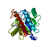
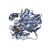
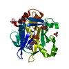
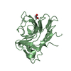
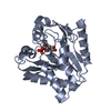
- PDB-4lru: Crystal structure of glyoxalase III (Orf 19.251) from Candida albicans -
+ Open data

- Basic information
| Entry | Database: PDB / ID: 4lru | ||||||
|---|---|---|---|---|---|---|---|
| Title | Crystal structure of glyoxalase III (Orf 19.251) from Candida albicans | ||||||
 Components Components | Glyoxalase III (glutathione-independent) | ||||||
 Keywords Keywords |  LYASE / DJ-1 superfamily LYASE / DJ-1 superfamily | ||||||
| Function / homology |  Function and homology informationD-lactate dehydratase / yeast-form cell wall / glyoxalase III activity / methylglyoxal catabolic process to D-lactate via S-lactoyl-glutathione / fungal biofilm matrix / hyphal cell wall Function and homology informationD-lactate dehydratase / yeast-form cell wall / glyoxalase III activity / methylglyoxal catabolic process to D-lactate via S-lactoyl-glutathione / fungal biofilm matrix / hyphal cell wallSimilarity search - Function | ||||||
| Biological species |  Candida albicans (yeast) Candida albicans (yeast) | ||||||
| Method | X-RAY DIFFRACTION / SYNCHROTRON / MOLECULAR REPLACEMENT / Resolution: 1.6 Å | ||||||
 Authors Authors | Hasim, S. / Hussin, N.A. / Nickerson, K.W. / Wilson, M.A. | ||||||
 Citation Citation | Journal: J.Biol.Chem. / Year: 2014 Title: A Glutathione-independent Glyoxalase of the DJ-1 Superfamily Plays an Important Role in Managing Metabolically Generated Methylglyoxal in Candida albicans. Authors: Hasim, S. / Hussin, N.A. / Alomar, F. / Bidasee, K.R. / Nickerson, K.W. / Wilson, M.A. | ||||||
| History |
|
- Structure visualization
| Structure viewer | Molecule: MolmilJmol/JSmol |
|---|
- Downloads & links
-Download
| PDBx/mmCIF format | 4lru.cif.gz | 113.8 KB | Display | PDBx/mmCIF format |
|---|---|---|---|---|
| PDB format | pdb4lru.ent.gz | 87.2 KB | Display | PDB format |
| PDBx/mmJSON format | 4lru.json.gz | Tree view | PDBx/mmJSON format | |
| Others |  Other downloads Other downloads |
-Validation report
| Arichive directory | https://data.pdbj.org/pub/pdb/validation_reports/lr/4lruftp://data.pdbj.org/pub/pdb/validation_reports/lr/4lru | HTTPS FTP |
|---|
-Related structure data
| Related structure data |  1rw7 S: Starting model for refinement |
|---|---|
| Similar structure data |

-Links
 PDBj
PDBj

- Assembly
| Deposited unit | 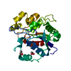
| ||||||||
|---|---|---|---|---|---|---|---|---|---|
| 1 |
| ||||||||
| Unit cell |
|
-Components
| #1: Protein | Mass: 26121.219 Da / Num. of mol.: 1 Source method: isolated from a genetically manipulated source Source: (gene. exp.) Candida albicans (yeast) / Strain: SC5314 / Gene: CaO19.7882, Orf 19.251, orf19.7882 / Plasmid: pET15b / Production host:  Escherichia coli (E. coli) / Strain (production host): BL21(DE3) / References: UniProt: Q5AF03, D-lactate dehydratase Escherichia coli (E. coli) / Strain (production host): BL21(DE3) / References: UniProt: Q5AF03, D-lactate dehydratase |
|---|---|
| #2: Chemical | ChemComp-EDO / Ethylene glycol  Mass: 62.068 Da / Num. of mol.: 1 / Source method: obtained synthetically / Formula: C2H6O2 Mass: 62.068 Da / Num. of mol.: 1 / Source method: obtained synthetically / Formula: C2H6O2 |
| #3: Chemical | ChemComp-ACT / Acetate  Mass: 59.044 Da / Num. of mol.: 1 / Source method: obtained synthetically / Formula: C2H3O2 Mass: 59.044 Da / Num. of mol.: 1 / Source method: obtained synthetically / Formula: C2H3O2 |
| #4: Water | ChemComp-HOH / Water Mass: 18.015 Da / Num. of mol.: 279 / Source method: isolated from a natural source / Formula: H2O Mass: 18.015 Da / Num. of mol.: 279 / Source method: isolated from a natural source / Formula: H2O |
-Experimental details
-Experiment
| Experiment | Method: X-RAY DIFFRACTION / Number of used crystals: 1 |
|---|
- Sample preparation
| Crystal | Density Matthews: 2.42 Å3/Da / Density % sol: 49.1 % |
|---|---|
| Crystal grow | Temperature: 277 K / pH: 4.1 Details: 100 mM sodium acetate pH 4.1, 120 mM ammonium acetate, 27% PEG4000 and 3% ethylene glycol, VAPOR DIFFUSION, SITTING DROP, temperature 277K |
-Data collection
| Diffraction | Mean temperature: 100 K |
|---|---|
| Diffraction source | Source: SYNCHROTRON / Site: SSRL  / Beamline: BL9-2 / Wavelength: 0.9 / Beamline: BL9-2 / Wavelength: 0.9 |
| Detector | Type: MARMOSAIC 325 mm CCD / Detector: CCD / Date: Apr 14, 2011 |
| Radiation | Monochromator: SI(111) DOUBLE CRYSTAL / Protocol: SINGLE WAVELENGTH / Monochromatic (M) / Laue (L): M / Scattering type: x-ray |
| Radiation wavelength | Wavelength: 0.9 Å / Relative weight: 1 |
| Reflection | Resolution: 1.6→41 Å / Num. obs: 34390 / % possible obs: 100 % / Observed criterion σ(I): 0 / Redundancy: 12.1 % / Biso Wilson estimate: 21 Å2 / Rmerge(I) obs: 0.05 / Net I/σ(I): 51.1 |
| Reflection shell | Resolution: 1.6→1.63 Å / Redundancy: 12.2 % / Rmerge(I) obs: 1 / Mean I/σ(I) obs: 2.2 / % possible all: 100 |
- Processing
| Software |
| ||||||||||||||||||||||||||||||||||||||||||||||||||||||||||||||||||||||||||||||||||||||||||||||||||||||||||||||||||||||||||||||||||||||||||||||||||||||||||||||||||||||||||||||||||||||
|---|---|---|---|---|---|---|---|---|---|---|---|---|---|---|---|---|---|---|---|---|---|---|---|---|---|---|---|---|---|---|---|---|---|---|---|---|---|---|---|---|---|---|---|---|---|---|---|---|---|---|---|---|---|---|---|---|---|---|---|---|---|---|---|---|---|---|---|---|---|---|---|---|---|---|---|---|---|---|---|---|---|---|---|---|---|---|---|---|---|---|---|---|---|---|---|---|---|---|---|---|---|---|---|---|---|---|---|---|---|---|---|---|---|---|---|---|---|---|---|---|---|---|---|---|---|---|---|---|---|---|---|---|---|---|---|---|---|---|---|---|---|---|---|---|---|---|---|---|---|---|---|---|---|---|---|---|---|---|---|---|---|---|---|---|---|---|---|---|---|---|---|---|---|---|---|---|---|---|---|---|---|---|---|
| Refinement | Method to determine structure: MOLECULAR REPLACEMENT Starting model: 1RW7 1rw7 Resolution: 1.6→41 Å / Cor.coef. Fo:Fc: 0.975 / Cor.coef. Fo:Fc free: 0.968 / SU B: 3.067 / SU ML: 0.054 / Cross valid method: THROUGHOUT / σ(F): 0 / ESU R: 0.078 / ESU R Free: 0.079 / Stereochemistry target values: MAXIMUM LIKELIHOOD / Details: HYDROGENS HAVE BEEN ADDED IN THE RIDING POSITIONS
| ||||||||||||||||||||||||||||||||||||||||||||||||||||||||||||||||||||||||||||||||||||||||||||||||||||||||||||||||||||||||||||||||||||||||||||||||||||||||||||||||||||||||||||||||||||||
| Solvent computation | Ion probe radii: 0.8 Å / Shrinkage radii: 0.8 Å / VDW probe radii: 1.2 Å / Solvent model: MASK | ||||||||||||||||||||||||||||||||||||||||||||||||||||||||||||||||||||||||||||||||||||||||||||||||||||||||||||||||||||||||||||||||||||||||||||||||||||||||||||||||||||||||||||||||||||||
| Displacement parameters | Biso mean: 25.58 Å2
| ||||||||||||||||||||||||||||||||||||||||||||||||||||||||||||||||||||||||||||||||||||||||||||||||||||||||||||||||||||||||||||||||||||||||||||||||||||||||||||||||||||||||||||||||||||||
| Refinement step | Cycle: LAST / Resolution: 1.6→41 Å
| ||||||||||||||||||||||||||||||||||||||||||||||||||||||||||||||||||||||||||||||||||||||||||||||||||||||||||||||||||||||||||||||||||||||||||||||||||||||||||||||||||||||||||||||||||||||
| Refine LS restraints |
|