 Movie
Movie Controller


+ Open data

- Basic information
| Entry | Database: PDB / ID: 4lpa | ||||||
|---|---|---|---|---|---|---|---|
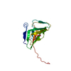
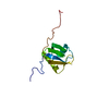
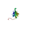
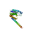
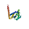
| Title | Crystal structure of a Cdc6 phosphopeptide in complex with Cks1 | ||||||
 Components Components | Cyclin-dependent kinases regulatory subunit | ||||||
 Keywords Keywords |  PROTEIN BINDING / TRANSFERASE REGULATOR / Phospho-protein PROTEIN BINDING / TRANSFERASE REGULATOR / Phospho-protein | ||||||
| Function / homology |  Function and homology information Function and homology informationregulation of cell cycle G1/S phase transition / cyclin-dependent protein serine/threonine kinase activator activity / SCF ubiquitin ligase complex / protein kinase activator activity / cyclin-dependent protein kinase holoenzyme complex / regulation of mitotic cell cycle / ubiquitin binding / histone binding / cell cycle / cell division ...regulation of cell cycle G1/S phase transition / cyclin-dependent protein serine/threonine kinase activator activity / SCF ubiquitin ligase complex / protein kinase activator activity / cyclin-dependent protein kinase holoenzyme complex / regulation of mitotic cell cycle / ubiquitin binding / histone binding / cell cycle / cell division / regulation of transcription by RNA polymerase II / protein kinase binding / positive regulation of transcription by RNA polymerase II / nucleus / cytoplasmSimilarity search - Function | ||||||
| Biological species |  Saccharomyces cerevisiae (brewer's yeast) Saccharomyces cerevisiae (brewer's yeast) | ||||||
| Method | X-RAY DIFFRACTION / SYNCHROTRON / MOLECULAR REPLACEMENT / Resolution: 2.9 Å | ||||||
 Authors Authors | McGrath, D.A. / Balog, E.R.M. / Koivomagi, M. / Lucena, R. / Mai, M.V. / Hirschi, A. / Kellogg, D.R. / Loog, M. / Rubin, S.M. | ||||||
 Citation Citation | Journal: Nat.Struct.Mol.Biol. / Year: 2013 Title: Cks confers specificity to phosphorylation-dependent CDK signaling pathways. Authors: McGrath, D.A. / Balog, E.R. / Koivomagi, M. / Lucena, R. / Mai, M.V. / Hirschi, A. / Kellogg, D.R. / Loog, M. / Rubin, S.M. | ||||||
| History |
|
- Structure visualization
| Structure viewer | Molecule: MolmilJmol/JSmol |
|---|
- Downloads & links
-Download
| PDBx/mmCIF format | 4lpa.cif.gz | 106.1 KB | Display | PDBx/mmCIF format |
|---|---|---|---|---|
| PDB format | pdb4lpa.ent.gz | 83.4 KB | Display | PDB format |
| PDBx/mmJSON format | 4lpa.json.gz | Tree view | PDBx/mmJSON format | |
| Others |  Other downloads Other downloads |
-Validation report
| Arichive directory | https://data.pdbj.org/pub/pdb/validation_reports/lp/4lpaftp://data.pdbj.org/pub/pdb/validation_reports/lp/4lpa | HTTPS FTP |
|---|
-Related structure data
| Related structure data |  1qb3S S: Starting model for refinement |
|---|---|
| Similar structure data |


-Links
 PDBj
PDBj- Assembly
| Deposited unit | 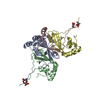
| ||||||||
|---|---|---|---|---|---|---|---|---|---|
| 1 | 
| ||||||||
| 2 | 
| ||||||||
| 3 | 
| ||||||||
| 4 | 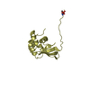
| ||||||||
| 5 | 
| ||||||||
| Unit cell |
|
-Components
| #1: Protein | Mass: 14261.974 Da / Num. of mol.: 4 Fragment: Cyclin-dependent kinases regulatory subunit (unp residues 1-113) Source method: isolated from a genetically manipulated source Source: (gene. exp.) Saccharomyces cerevisiae (brewer's yeast)Strain: ATCC 204508 / S288c / Gene: CKS1, YBR1011, YBR135W / Plasmid: pET / Production host:  Escherichia coli (E. coli) / Strain (production host): BL21(DE3) / References: UniProt: P20486 Escherichia coli (E. coli) / Strain (production host): BL21(DE3) / References: UniProt: P20486#2: Water | ChemComp-HOH / | Water Mass: 18.015 Da / Num. of mol.: 52 / Source method: isolated from a natural source / Formula: H2O Mass: 18.015 Da / Num. of mol.: 52 / Source method: isolated from a natural source / Formula: H2O |
|---|
-Experimental details
-Experiment
| Experiment | Method: X-RAY DIFFRACTION / Number of used crystals: 1 |
|---|
- Sample preparation
| Crystal | Density Matthews: 3.77 Å3/Da / Density % sol: 67.38 % |
|---|---|
| Crystal grow | Temperature: 293.15 K / Method: vapor diffusion, sitting drop / pH: 5.5 Details: 400mM potassium sodium tartrate, 0.1M MES, pH 5.5, VAPOR DIFFUSION, SITTING DROP, temperature 293.15K |
-Data collection
| Diffraction | Mean temperature: 100 K |
|---|---|
| Diffraction source | Source: SYNCHROTRON / Site: APS  / Beamline: 23-ID-D / Wavelength: 1.0332 Å / Beamline: 23-ID-D / Wavelength: 1.0332 Å |
| Detector | Type: MARMOSAIC 300 mm CCD / Detector: CCD / Date: Jul 5, 2012 |
| Radiation | Monochromator: Si(111) / Protocol: SINGLE WAVELENGTH / Monochromatic (M) / Laue (L): M / Scattering type: x-ray |
| Radiation wavelength | Wavelength: 1.0332 Å / Relative weight: 1 |
| Reflection | Resolution: 2.9→59.98 Å / Num. all: 20290 / Num. obs: 20254 / % possible obs: 99.79 % / Observed criterion σ(F): 0 / Observed criterion σ(I): -3 / Rmerge(I) obs: 0.0463 / Net I/σ(I): 12.65 |
| Reflection shell | Resolution: 2.9→3.004 Å / Rmerge(I) obs: 0.2598 / Mean I/σ(I) obs: 3.57 / % possible all: 99.9 |
- Processing
| Software |
| |||||||||||||||||||||||||||||||||||||||||||||||||||||||||||||||||||||||||||||||||||||||||||||||||||||||||
|---|---|---|---|---|---|---|---|---|---|---|---|---|---|---|---|---|---|---|---|---|---|---|---|---|---|---|---|---|---|---|---|---|---|---|---|---|---|---|---|---|---|---|---|---|---|---|---|---|---|---|---|---|---|---|---|---|---|---|---|---|---|---|---|---|---|---|---|---|---|---|---|---|---|---|---|---|---|---|---|---|---|---|---|---|---|---|---|---|---|---|---|---|---|---|---|---|---|---|---|---|---|---|---|---|---|---|
| Refinement | Method to determine structure: MOLECULAR REPLACEMENT Starting model: PDB ENTRY 1QB3 Resolution: 2.9→58.147 Å / SU ML: 0.43 / σ(F): 1.36 / Phase error: 31.86 / Stereochemistry target values: MLHL
| |||||||||||||||||||||||||||||||||||||||||||||||||||||||||||||||||||||||||||||||||||||||||||||||||||||||||
| Solvent computation | Shrinkage radii: 0.9 Å / VDW probe radii: 1.11 Å / Solvent model: FLAT BULK SOLVENT MODEL | |||||||||||||||||||||||||||||||||||||||||||||||||||||||||||||||||||||||||||||||||||||||||||||||||||||||||
| Refinement step | Cycle: LAST / Resolution: 2.9→58.147 Å
| |||||||||||||||||||||||||||||||||||||||||||||||||||||||||||||||||||||||||||||||||||||||||||||||||||||||||
| Refine LS restraints |
| |||||||||||||||||||||||||||||||||||||||||||||||||||||||||||||||||||||||||||||||||||||||||||||||||||||||||
| LS refinement shell |
|
