 Movie
Movie Controller

[English] 日本語
 Yorodumi
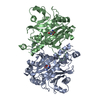
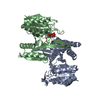
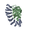
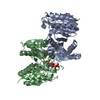
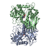
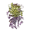
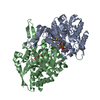
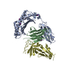
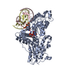
- PDB-4lns: Crystal structure of Asparagine synthetase A (AsnA) from Trypanos... -
+ Open data

- Basic information
| Entry | Database: PDB / ID: 4lns | ||||||
|---|---|---|---|---|---|---|---|
| Title | Crystal structure of Asparagine synthetase A (AsnA) from Trypanosoma brucei | ||||||
 Components Components | Asparagine synthetase a | ||||||
 Keywords Keywords |  LIGASE / Asparagine synthetase A LIGASE / Asparagine synthetase A | ||||||
| Function / homology |  Function and homology informationaspartate-ammonia ligase / aspartate-ammonia ligase activity / asparagine biosynthetic process / cytosol Function and homology informationaspartate-ammonia ligase / aspartate-ammonia ligase activity / asparagine biosynthetic process / cytosolSimilarity search - Function | ||||||
| Biological species |  Trypanosoma brucei brucei (eukaryote) Trypanosoma brucei brucei (eukaryote) | ||||||
| Method | X-RAY DIFFRACTION / SYNCHROTRON / MOLECULAR REPLACEMENT / Resolution: 2.2 Å | ||||||
 Authors Authors | Khan, S. / Madhubala, R. / Sharma, A. | ||||||
 Citation Citation | Journal: J.Biol.Chem. / Year: 2014 Title: Identification and functional characterization of a novel bacterial type asparagine synthetase A: a tRNA synthetase paralog from Leishmania donovani. Authors: Manhas, R. / Tripathi, P. / Khan, S. / Sethu Lakshmi, B. / Lal, S.K. / Gowri, V.S. / Sharma, A. / Madhubala, R. | ||||||
| History |
|
- Structure visualization
| Structure viewer | Molecule: MolmilJmol/JSmol |
|---|
- Downloads & links
-Download
| PDBx/mmCIF format | 4lns.cif.gz | 75 KB | Display | PDBx/mmCIF format |
|---|---|---|---|---|
| PDB format | pdb4lns.ent.gz | 56.5 KB | Display | PDB format |
| PDBx/mmJSON format | 4lns.json.gz | Tree view | PDBx/mmJSON format | |
| Others |  Other downloads Other downloads |
-Validation report
| Arichive directory | https://data.pdbj.org/pub/pdb/validation_reports/ln/4lnsftp://data.pdbj.org/pub/pdb/validation_reports/ln/4lns | HTTPS FTP |
|---|
-Related structure data
| Related structure data | |
|---|---|
| Similar structure data |


-Links
 PDBj
PDBj
- Assembly
| Deposited unit | 
| ||||||||
|---|---|---|---|---|---|---|---|---|---|
| 1 | 
| ||||||||
| Unit cell |
|
-Components
| #1: Protein | Mass: 39649.344 Da / Num. of mol.: 1 Source method: isolated from a genetically manipulated source Source: (gene. exp.) Trypanosoma brucei brucei (eukaryote) / Strain: 927/4 GUTat10.1 / Gene: Tb927.7.1110 / Production host:  Escherichia coli (E. coli) / References: UniProt: Q57WT9, aspartate-ammonia ligase Escherichia coli (E. coli) / References: UniProt: Q57WT9, aspartate-ammonia ligase |
|---|---|
| #2: Water | ChemComp-HOH / Water Mass: 18.015 Da / Num. of mol.: 137 / Source method: isolated from a natural source / Formula: H2O Mass: 18.015 Da / Num. of mol.: 137 / Source method: isolated from a natural source / Formula: H2O |
-Experimental details
-Experiment
| Experiment | Method: X-RAY DIFFRACTION / Number of used crystals: 1 |
|---|
- Sample preparation
| Crystal | Density Matthews: 2.5 Å3/Da / Density % sol: 50.86 % |
|---|---|
| Crystal grow | Temperature: 293 K / Method: vapor diffusion, hanging drop / pH: 7.5 Details: 10% w/v PEG 20000, 20% v/v PEG MME 550, 0.03M of each NPS (sodium nitrate, disodium hydrogen phosphate, ammonium sulfate), 0.1M MOPS/HEPES-Na, pH 7.5, VAPOR DIFFUSION, HANGING DROP, temperature 293K |
-Data collection
| Diffraction | Mean temperature: 100 K |
|---|---|
| Diffraction source | Source: SYNCHROTRON / Site: ESRF  / Beamline: BM14 / Wavelength: 0.976 Å / Beamline: BM14 / Wavelength: 0.976 Å |
| Detector | Type: MARMOSAIC 225 mm CCD / Detector: CCD / Date: May 7, 2013 |
| Radiation | Monochromator: Si 111 CHANNEL / Protocol: SINGLE WAVELENGTH / Monochromatic (M) / Laue (L): M / Scattering type: x-ray |
| Radiation wavelength | Wavelength: 0.976 Å / Relative weight: 1 |
| Reflection | Resolution: 2.2→25 Å / Num. obs: 21721 / % possible obs: 99.6 % / Observed criterion σ(F): 0 / Observed criterion σ(I): 0 |
| Reflection shell | Resolution: 2.2→2.24 Å / Rmerge(I) obs: 0.36 / Mean I/σ(I) obs: 2.1 / % possible all: 100 |
- Processing
| Software |
| ||||||||||||||||||||||||||||||||||||||||||||||||||||||
|---|---|---|---|---|---|---|---|---|---|---|---|---|---|---|---|---|---|---|---|---|---|---|---|---|---|---|---|---|---|---|---|---|---|---|---|---|---|---|---|---|---|---|---|---|---|---|---|---|---|---|---|---|---|---|---|
| Refinement | Method to determine structure: MOLECULAR REPLACEMENT / Resolution: 2.2→24.647 Å / Occupancy max: 1 / Occupancy min: 1 / FOM work R set: 0.8257 / SU ML: 0.59 / σ(F): 1.34 / Phase error: 23.06 / Stereochemistry target values: ML
| ||||||||||||||||||||||||||||||||||||||||||||||||||||||
| Solvent computation | Shrinkage radii: 0.83 Å / VDW probe radii: 1.1 Å / Solvent model: FLAT BULK SOLVENT MODEL / Bsol: 61.433 Å2 / ksol: 0.4 e/Å3 | ||||||||||||||||||||||||||||||||||||||||||||||||||||||
| Displacement parameters | Biso max: 101.2 Å2 / Biso mean: 40.4514 Å2 / Biso min: 20.11 Å2
| ||||||||||||||||||||||||||||||||||||||||||||||||||||||
| Refinement step | Cycle: LAST / Resolution: 2.2→24.647 Å
| ||||||||||||||||||||||||||||||||||||||||||||||||||||||
| Refine LS restraints |
| ||||||||||||||||||||||||||||||||||||||||||||||||||||||
| LS refinement shell | Refine-ID: X-RAY DIFFRACTION / Total num. of bins used: 8
|
