 Movie
Movie Controller

[English] 日本語
 Yorodumi
- PDB-4ln2: The second SH3 domain from CAP/Ponsin in complex with proline ric... -
+ Open data

- Basic information
| Entry | Database: PDB / ID: 4ln2 | ||||||
|---|---|---|---|---|---|---|---|
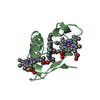
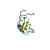
| Title | The second SH3 domain from CAP/Ponsin in complex with proline rich peptide from Vinculin | ||||||
 Components Components |
| ||||||
 Keywords Keywords |  SIGNALING PROTEIN / sh3 domain / cell migration / focal adhesion SIGNALING PROTEIN / sh3 domain / cell migration / focal adhesion | ||||||
| Function / homology |  Function and homology information Function and homology informationregulation of protein localization to adherens junction / podosome ring / outer dense plaque of desmosome / inner dense plaque of desmosome / terminal web / cell-substrate junction / epithelial cell-cell adhesion / zonula adherens / dystroglycan binding / alpha-catenin binding ...regulation of protein localization to adherens junction / podosome ring / outer dense plaque of desmosome / inner dense plaque of desmosome / terminal web / cell-substrate junction / epithelial cell-cell adhesion / zonula adherens / dystroglycan binding / alpha-catenin binding / flotillin complex / fascia adherens / cell-cell contact zone / apical junction assembly / costamere / regulation of establishment of endothelial barrier / adherens junction assembly / focal adhesion assembly / axon extension / protein localization to cell surface / lamellipodium assembly / regulation of focal adhesion assembly / maintenance of blood-brain barrier / stress fiber assembly / cell-substrate adhesion / brush border / Signaling by ALK fusions and activated point mutants / Smooth Muscle Contraction / positive regulation of glycogen biosynthetic process / positive regulation of lipid biosynthetic process / stress fiber / positive regulation of insulin receptor signaling pathway / cytoskeletal protein binding / cell-matrix adhesion / negative regulation of cell migration / cell projection / morphogenesis of an epithelium / positive regulation of protein localization to plasma membrane / positive regulation of glucose import / adherens junction / Signaling by high-kinase activity BRAF mutants / sarcolemma / MAP2K and MAPK activation / insulin receptor binding / platelet aggregation / beta-catenin binding / nuclear matrix / cellular response to insulin stimulus / specific granule lumen / Signaling by RAF1 mutants / Signaling by moderate kinase activity BRAF mutants / Paradoxical activation of RAF signaling by kinase inactive BRAF / Signaling downstream of RAS mutants / extracellular vesicle / Signaling by BRAF and RAF1 fusions / cell-cell junction / signaling receptor complex adaptor activity / Platelet degranulation / insulin receptor signaling pathway / actin binding / secretory granule lumen / ficolin-1-rich granule lumen / molecular adaptor activity / cytoskeleton / cell adhesion / cadherin binding / membrane raft / focal adhesion / centrosome / ubiquitin protein ligase binding / Neutrophil degranulation / structural molecule activity / protein-containing complex / extracellular exosome / extracellular region / nucleus / plasma membrane / cytosol / cytoplasmSimilarity search - Function | ||||||
| Biological species |  Homo sapiens (human) Homo sapiens (human) | ||||||
| Method | X-RAY DIFFRACTION / SYNCHROTRON / MOLECULAR REPLACEMENT / Resolution: 1 Å | ||||||
 Authors Authors | Zhao, D. / Li, F. / Wu, J. / Shi, Y. / Zhang, Z. / Gong, Q. | ||||||
 Citation Citation | Journal: J.Struct.Biol. / Year: 2014 Title: Structural investigation of the interaction between the tandem SH3 domains of c-Cbl-associated protein and vinculin Authors: Zhao, D. / Wang, X. / Peng, J. / Wang, C. / Li, F. / Sun, Q. / Zhang, Y. / Zhang, J. / Cai, G. / Zuo, X. / Wu, J. / Shi, Y. / Zhang, Z. / Gong, Q. | ||||||
| History |
|
- Structure visualization
| Structure viewer | Molecule: MolmilJmol/JSmol |
|---|
- Downloads & links
-Download
| PDBx/mmCIF format | 4ln2.cif.gz | 49.9 KB | Display | PDBx/mmCIF format |
|---|---|---|---|---|
| PDB format | pdb4ln2.ent.gz | 35.1 KB | Display | PDB format |
| PDBx/mmJSON format | 4ln2.json.gz | Tree view | PDBx/mmJSON format | |
| Others |  Other downloads Other downloads |
-Validation report
| Arichive directory | https://data.pdbj.org/pub/pdb/validation_reports/ln/4ln2ftp://data.pdbj.org/pub/pdb/validation_reports/ln/4ln2 | HTTPS FTP |
|---|
-Related structure data
| Related structure data |  2moxC  4lnpC  2o9sS C: citing same article ( S: Starting model for refinement |
|---|---|
| Similar structure data |


-Links
 PDBj
PDBj


- Assembly
| Deposited unit | 
| ||||||||
|---|---|---|---|---|---|---|---|---|---|
| 1 |
| ||||||||
| Unit cell |
|
-Components
| #1: Protein | Mass: 7970.064 Da / Num. of mol.: 1 / Fragment: UNP residues 866-930 Source method: isolated from a genetically manipulated source Source: (gene. exp.) Homo sapiens (human) / Gene: KIAA0894, KIAA1296, SH3D5, SORBS1 / Production host:  Escherichia coli (E. coli) / References: UniProt: Q9BX66 Escherichia coli (E. coli) / References: UniProt: Q9BX66 |
|---|---|
| #2: Protein/peptide | Mass: 1188.390 Da / Num. of mol.: 1 / Fragment: UNP residues 857-867 Source method: isolated from a genetically manipulated source Source: (gene. exp.) Homo sapiens (human) / Gene: VCL / Production host: Escherichia coli (E. coli) / References: UniProt: P18206 |
| #3: Water | ChemComp-HOH / Water Mass: 18.015 Da / Num. of mol.: 121 / Source method: isolated from a natural source / Formula: H2O Mass: 18.015 Da / Num. of mol.: 121 / Source method: isolated from a natural source / Formula: H2O |
-Experimental details
-Experiment
| Experiment | Method: X-RAY DIFFRACTION / Number of used crystals: 1 |
|---|
- Sample preparation
| Crystal | Density Matthews: 1.91 Å3/Da / Density % sol: 35.6 % |
|---|---|
| Crystal grow | Method: evaporation / Details: EVAPORATION |
-Data collection
| Diffraction | Mean temperature: 77 K |
|---|---|
| Diffraction source | Source: SYNCHROTRON / Site: SSRF  / Beamline: BL17U / Beamline: BL17U |
| Detector | Type: ADSC QUANTUM 315r / Detector: CCD / Date: May 7, 2012 |
| Radiation | Protocol: SINGLE WAVELENGTH / Monochromatic (M) / Laue (L): M / Scattering type: x-ray |
| Radiation wavelength | Relative weight: 1 |
| Reflection | Resolution: 1→34.64 Å / Num. all: 38774 / Num. obs: 36766 |
- Processing
| Software |
| ||||||||||||||||||||||||||||||||||||||||||||||||||||||||||||||||||||||||||||||||||||||||||||||||||||||||||||||||||||||||||||||||||||||||||||||||||||||||||||||||||||||||||||||||||||||
|---|---|---|---|---|---|---|---|---|---|---|---|---|---|---|---|---|---|---|---|---|---|---|---|---|---|---|---|---|---|---|---|---|---|---|---|---|---|---|---|---|---|---|---|---|---|---|---|---|---|---|---|---|---|---|---|---|---|---|---|---|---|---|---|---|---|---|---|---|---|---|---|---|---|---|---|---|---|---|---|---|---|---|---|---|---|---|---|---|---|---|---|---|---|---|---|---|---|---|---|---|---|---|---|---|---|---|---|---|---|---|---|---|---|---|---|---|---|---|---|---|---|---|---|---|---|---|---|---|---|---|---|---|---|---|---|---|---|---|---|---|---|---|---|---|---|---|---|---|---|---|---|---|---|---|---|---|---|---|---|---|---|---|---|---|---|---|---|---|---|---|---|---|---|---|---|---|---|---|---|---|---|---|---|
| Refinement | Method to determine structure: MOLECULAR REPLACEMENT Starting model: 2o9S Resolution: 1→26.43 Å / Cor.coef. Fo:Fc: 0.973 / Cor.coef. Fo:Fc free: 0.97 / SU B: 0.551 / SU ML: 0.014 / Cross valid method: THROUGHOUT / ESU R: 0.023 / ESU R Free: 0.023 / Stereochemistry target values: MAXIMUM LIKELIHOOD / Details: HYDROGENS HAVE BEEN ADDED IN THE RIDING POSITIONS
| ||||||||||||||||||||||||||||||||||||||||||||||||||||||||||||||||||||||||||||||||||||||||||||||||||||||||||||||||||||||||||||||||||||||||||||||||||||||||||||||||||||||||||||||||||||||
| Solvent computation | Ion probe radii: 0.8 Å / Shrinkage radii: 0.8 Å / VDW probe radii: 1.2 Å / Solvent model: MASK | ||||||||||||||||||||||||||||||||||||||||||||||||||||||||||||||||||||||||||||||||||||||||||||||||||||||||||||||||||||||||||||||||||||||||||||||||||||||||||||||||||||||||||||||||||||||
| Displacement parameters | Biso mean: 9.226 Å2
| ||||||||||||||||||||||||||||||||||||||||||||||||||||||||||||||||||||||||||||||||||||||||||||||||||||||||||||||||||||||||||||||||||||||||||||||||||||||||||||||||||||||||||||||||||||||
| Refinement step | Cycle: LAST / Resolution: 1→26.43 Å
| ||||||||||||||||||||||||||||||||||||||||||||||||||||||||||||||||||||||||||||||||||||||||||||||||||||||||||||||||||||||||||||||||||||||||||||||||||||||||||||||||||||||||||||||||||||||
| Refine LS restraints |
|