 Movie
Movie Controller

[English] 日本語
 Yorodumi
- PDB-4lks: Structure of CBM32-3 from a family 31 glycoside hydrolase from Cl... -
+ Open data

- Basic information
| Entry | Database: PDB / ID: 4lks | ||||||
|---|---|---|---|---|---|---|---|
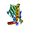
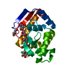
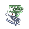
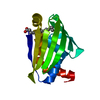
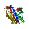
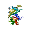
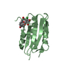
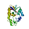
| Title | Structure of CBM32-3 from a family 31 glycoside hydrolase from Clostridium perfringens in complex with galactose | ||||||
 Components Components | Glycosyl hydrolase, family 31/fibronectin type III domain protein Glycoside hydrolase Glycoside hydrolase | ||||||
 Keywords Keywords | SUGAR BINDING PROTEIN / B-sandwich / carbohydrate-binding / galactose | ||||||
| Function / homology |  Function and homology information Function and homology informationpolysaccharide catabolic process / hydrolase activity, hydrolyzing O-glycosyl compounds / carbohydrate binding / metal ion bindingSimilarity search - Function | ||||||
| Biological species |   Clostridium perfringens (bacteria) Clostridium perfringens (bacteria) | ||||||
| Method | X-RAY DIFFRACTION / SYNCHROTRON / MOLECULAR REPLACEMENT / Resolution: 1.5 Å | ||||||
 Authors Authors | Grondin, J.M. / Duan, D. / Kirlin, A.C. / Furness, H.S. / Allingham, J.S. / Smith, S.P. | ||||||
 Citation Citation | Journal: Plos One / Year: 2017 Title: Diverse modes of galacto-specific carbohydrate recognition by a family 31 glycoside hydrolase from Clostridium perfringens. Authors: Grondin, J.M. / Duan, D. / Kirlin, A.C. / Abe, K.T. / Chitayat, S. / Spencer, H.L. / Spencer, C. / Campigotto, A. / Houliston, S. / Arrowsmith, C.H. / Allingham, J.S. / Boraston, A.B. / Smith, S.P. | ||||||
| History |
|
- Structure visualization
| Structure viewer | Molecule: MolmilJmol/JSmol |
|---|
- Downloads & links
-Download
| PDBx/mmCIF format | 4lks.cif.gz | 90.2 KB | Display | PDBx/mmCIF format |
|---|---|---|---|---|
| PDB format | pdb4lks.ent.gz | 66 KB | Display | PDB format |
| PDBx/mmJSON format | 4lks.json.gz | Tree view | PDBx/mmJSON format | |
| Others |  Other downloads Other downloads |
-Validation report
| Arichive directory | https://data.pdbj.org/pub/pdb/validation_reports/lk/4lksftp://data.pdbj.org/pub/pdb/validation_reports/lk/4lks | HTTPS FTP |
|---|
-Related structure data
| Related structure data | 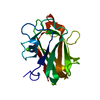 4lplC 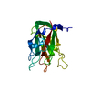 4lqrC 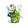 4p5yC 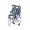 4uapC 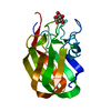 2j1aS C: citing same article ( S: Starting model for refinement |
|---|---|
| Similar structure data |

-Links
 PDBj
PDBj


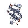
- Assembly
| Deposited unit | 
| ||||||||
|---|---|---|---|---|---|---|---|---|---|
| 1 | 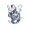
| ||||||||
| 2 | 
| ||||||||
| Unit cell |
|
-Components
| #1: Protein | Glycoside hydrolase Mass: 18993.844 Da / Num. of mol.: 2 / Fragment: CBM32-3, UNP residues 1640-1785 Source method: isolated from a genetically manipulated source Source: (gene. exp.) Clostridium perfringens (bacteria) / Strain: ATCC 13124 / NCTC 8237 / Type A / Gene: CPF_1301 / Production host: Escherichia coli (E. coli) / References: UniProt: Q0TRJ3, UniProt: A0A0H2YST8*PLUS#2: Chemical |   Mass: 40.078 Da / Num. of mol.: 2 / Source method: obtained synthetically / Formula: Ca Mass: 40.078 Da / Num. of mol.: 2 / Source method: obtained synthetically / Formula: Ca#3: Sugar | Galactose  Type: D-saccharide, beta linking / Mass: 180.156 Da / Num. of mol.: 2 Type: D-saccharide, beta linking / Mass: 180.156 Da / Num. of mol.: 2Source method: isolated from a genetically manipulated source Formula: C6H12O6 #4: Water | ChemComp-HOH / | Water Mass: 18.015 Da / Num. of mol.: 515 / Source method: isolated from a natural source / Formula: H2O Mass: 18.015 Da / Num. of mol.: 515 / Source method: isolated from a natural source / Formula: H2O |
|---|
-Experimental details
-Experiment
| Experiment | Method: X-RAY DIFFRACTION / Number of used crystals: 1 |
|---|
- Sample preparation
| Crystal | Density Matthews: 1.95 Å3/Da / Density % sol: 36.3 % |
|---|---|
| Crystal grow | Temperature: 298 K / Method: vapor diffusion, hanging drop / pH: 8 Details: 1.6M ammonium citrate, pH 8.0, VAPOR DIFFUSION, HANGING DROP, temperature 298K |
-Data collection
| Diffraction | Mean temperature: 93.15 K | |||||||||||||||||||||||||||||||||||||||||||||||||||||||||||||||||||||||||||||
|---|---|---|---|---|---|---|---|---|---|---|---|---|---|---|---|---|---|---|---|---|---|---|---|---|---|---|---|---|---|---|---|---|---|---|---|---|---|---|---|---|---|---|---|---|---|---|---|---|---|---|---|---|---|---|---|---|---|---|---|---|---|---|---|---|---|---|---|---|---|---|---|---|---|---|---|---|---|---|
| Diffraction source | Source: SYNCHROTRON / Site: NSLS  / Beamline: X6A / Wavelength: 0.98 Å / Beamline: X6A / Wavelength: 0.98 Å | |||||||||||||||||||||||||||||||||||||||||||||||||||||||||||||||||||||||||||||
| Detector | Type: ADSC QUANTUM 210 / Detector: CCD / Date: Jul 13, 2011 | |||||||||||||||||||||||||||||||||||||||||||||||||||||||||||||||||||||||||||||
| Radiation | Monochromator: Si (III) channel cut / Protocol: SINGLE WAVELENGTH / Monochromatic (M) / Laue (L): M / Scattering type: x-ray | |||||||||||||||||||||||||||||||||||||||||||||||||||||||||||||||||||||||||||||
| Radiation wavelength | Wavelength: 0.98 Å / Relative weight: 1 | |||||||||||||||||||||||||||||||||||||||||||||||||||||||||||||||||||||||||||||
| Reflection | Resolution: 1.5→40 Å / Num. obs: 53421 / % possible obs: 98.9 % / Redundancy: 4.2 % / Rmerge(I) obs: 0.051 / Χ2: 1.236 / Net I/σ(I): 16.4 | |||||||||||||||||||||||||||||||||||||||||||||||||||||||||||||||||||||||||||||
| Reflection shell |
|
- Processing
| Software |
| ||||||||||||||||||||||||||||||||||||||||||||||||||||||||||||||||||||||||||||||||||||||||||||||||||||||||||||||||||||||||||||||||||||||||||||
|---|---|---|---|---|---|---|---|---|---|---|---|---|---|---|---|---|---|---|---|---|---|---|---|---|---|---|---|---|---|---|---|---|---|---|---|---|---|---|---|---|---|---|---|---|---|---|---|---|---|---|---|---|---|---|---|---|---|---|---|---|---|---|---|---|---|---|---|---|---|---|---|---|---|---|---|---|---|---|---|---|---|---|---|---|---|---|---|---|---|---|---|---|---|---|---|---|---|---|---|---|---|---|---|---|---|---|---|---|---|---|---|---|---|---|---|---|---|---|---|---|---|---|---|---|---|---|---|---|---|---|---|---|---|---|---|---|---|---|---|---|---|
| Refinement | Method to determine structure: MOLECULAR REPLACEMENT Starting model: PDB ENTRY 2J1A Resolution: 1.5→30.659 Å / Occupancy max: 1 / Occupancy min: 0.31 / FOM work R set: 0.8808 / SU ML: 0.19 / σ(F): 1.34 / Phase error: 18.55 / Stereochemistry target values: ML
| ||||||||||||||||||||||||||||||||||||||||||||||||||||||||||||||||||||||||||||||||||||||||||||||||||||||||||||||||||||||||||||||||||||||||||||
| Solvent computation | Shrinkage radii: 0.73 Å / VDW probe radii: 1 Å / Solvent model: FLAT BULK SOLVENT MODEL / Bsol: 39.324 Å2 / ksol: 0.394 e/Å3 | ||||||||||||||||||||||||||||||||||||||||||||||||||||||||||||||||||||||||||||||||||||||||||||||||||||||||||||||||||||||||||||||||||||||||||||
| Displacement parameters | Biso max: 69.66 Å2 / Biso mean: 16.3428 Å2 / Biso min: 5.62 Å2
| ||||||||||||||||||||||||||||||||||||||||||||||||||||||||||||||||||||||||||||||||||||||||||||||||||||||||||||||||||||||||||||||||||||||||||||
| Refinement step | Cycle: LAST / Resolution: 1.5→30.659 Å
| ||||||||||||||||||||||||||||||||||||||||||||||||||||||||||||||||||||||||||||||||||||||||||||||||||||||||||||||||||||||||||||||||||||||||||||
| Refine LS restraints |
| ||||||||||||||||||||||||||||||||||||||||||||||||||||||||||||||||||||||||||||||||||||||||||||||||||||||||||||||||||||||||||||||||||||||||||||
| LS refinement shell | Refine-ID: X-RAY DIFFRACTION / Total num. of bins used: 19
|
