 Movie
Movie Controller

[English] 日本語
 Yorodumi
- PDB-4lkd: Crystal Structure of Pseudomonas aeruginosa Lectin LecA Complexed... -
+ Open data

- Basic information
| Entry | Database: PDB / ID: 4lkd | ||||||
|---|---|---|---|---|---|---|---|
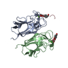
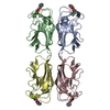
| Title | Crystal Structure of Pseudomonas aeruginosa Lectin LecA Complexed with GalA-QRS at 2.31 A Resolution | ||||||
 Components Components |
| ||||||
 Keywords Keywords | SUGAR BINDING PROTEIN/INHIBITOR / Lectin Fold / Sugar Binding Protein /  Galactose / SUGAR BINDING PROTEIN-INHIBITOR complex Galactose / SUGAR BINDING PROTEIN-INHIBITOR complex | ||||||
| Function / homology |  Function and homology information Function and homology informationheterophilic cell-cell adhesion via plasma membrane cell adhesion molecules / carbohydrate binding / periplasmic space / cell surface / cytoplasmSimilarity search - Function | ||||||
| Biological species |   Pseudomonas aeruginosa (bacteria) Pseudomonas aeruginosa (bacteria) | ||||||
| Method | X-RAY DIFFRACTION / SYNCHROTRON / MOLECULAR REPLACEMENT / Resolution: 2.307 Å | ||||||
 Authors Authors | Kadam, R.U. / Stocker, A. / Reymond, J.-L. | ||||||
 Citation Citation | Journal: Chemistry / Year: 2013 Title: Structure-Based Optimization of the Terminal Tripeptide in Glycopeptide Dendrimer Inhibitors of Pseudomonas aeruginosa Biofilms Targeting LecA. Authors: Kadam, R.U. / Bergmann, M. / Garg, D. / Gabrieli, G. / Stocker, A. / Darbre, T. / Reymond, J.-L. | ||||||
| History |
|
- Structure visualization
| Structure viewer | Molecule: MolmilJmol/JSmol |
|---|
- Downloads & links
-Download
| PDBx/mmCIF format | 4lkd.cif.gz | 220.7 KB | Display | PDBx/mmCIF format |
|---|---|---|---|---|
| PDB format | pdb4lkd.ent.gz | 179.7 KB | Display | PDB format |
| PDBx/mmJSON format | 4lkd.json.gz | Tree view | PDBx/mmJSON format | |
| Others |  Other downloads Other downloads |
-Validation report
| Arichive directory | https://data.pdbj.org/pub/pdb/validation_reports/lk/4lkdftp://data.pdbj.org/pub/pdb/validation_reports/lk/4lkd | HTTPS FTP |
|---|
-Related structure data


-Links
 PDBj
PDBj

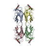
- Assembly
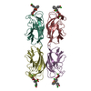
| Deposited unit | 
| |||||||||
|---|---|---|---|---|---|---|---|---|---|---|
| 1 | 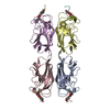
| |||||||||
| 2 | 
| |||||||||
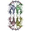
| Unit cell |
| |||||||||
| Components on special symmetry positions |
|
-Components
-Protein / Protein/peptide / Sugars , 3 types, 24 molecules ABCDEFGHIJKLMNOP
| #1: Protein | Mass: 12770.137 Da / Num. of mol.: 8 Source method: isolated from a genetically manipulated source Source: (gene. exp.) Pseudomonas aeruginosa (bacteria) / Strain: ATCC 15692 / PAO1 / 1C / PRS 101 / LMG 12228 / Gene: lecA, pa1L, PA2570 / Plasmid: PET25PAIL / Production host: Escherichia coli (E. coli) / Strain (production host): Bl21(DE3) / References: UniProt: Q05097#2: Protein/peptide | Mass: 461.493 Da / Num. of mol.: 8 / Source method: obtained synthetically / Details: The peptide was chemically synthesized. #3: Sugar | ChemComp-GAL / Galactose Type: D-saccharide, beta linking / Mass: 180.156 Da / Num. of mol.: 8 Type: D-saccharide, beta linking / Mass: 180.156 Da / Num. of mol.: 8Source method: isolated from a genetically manipulated source Formula: C6H12O6 |
|---|
-Non-polymers , 3 types, 1152 molecules 

| #4: Chemical | ChemComp-CA /  Mass: 40.078 Da / Num. of mol.: 8 / Source method: obtained synthetically / Formula: Ca Mass: 40.078 Da / Num. of mol.: 8 / Source method: obtained synthetically / Formula: Ca#5: Chemical | ChemComp-PHB / 4-Hydroxybenzoic acid Mass: 138.121 Da / Num. of mol.: 8 / Source method: obtained synthetically / Formula: C7H6O3 Mass: 138.121 Da / Num. of mol.: 8 / Source method: obtained synthetically / Formula: C7H6O3#6: Water | ChemComp-HOH / | WaterMass: 18.015 Da / Num. of mol.: 1136 / Source method: isolated from a natural source / Formula: H2O |
|---|
-Experimental details
-Experiment
| Experiment | Method: X-RAY DIFFRACTION / Number of used crystals: 1 |
|---|
- Sample preparation
| Crystal | Density Matthews: 4.56 Å3/Da / Density % sol: 73.03 % Description: THE ENTRY CONTAINS FRIEDEL PAIRS IN F_PLUS/MINUS COLUMNS |
|---|---|
| Crystal grow | Temperature: 293 K / Method: vapor diffusion, sitting drop / pH: 8.5 Details: 1.5M Lithium sulfate monohydrate, 0.1M Tris pH 8.5 , VAPOR DIFFUSION, SITTING DROP, temperature 293K |
-Data collection
| Diffraction | Mean temperature: 100 K |
|---|---|
| Diffraction source | Source: SYNCHROTRON / Site: SLS  / Beamline: X06DA / Wavelength: 1 Å / Beamline: X06DA / Wavelength: 1 Å |
| Detector | Type: MARMOSAIC 225 mm CCD / Detector: CCD / Date: May 14, 2011 |
| Radiation | Monochromator: Bartels Monochromator / Protocol: SINGLE WAVELENGTH / Monochromatic (M) / Laue (L): M / Scattering type: x-ray |
| Radiation wavelength | Wavelength: 1 Å / Relative weight: 1 |
| Reflection | Resolution: 2.307→48.461 Å / Num. all: 81750 / Num. obs: 81750 / % possible obs: 98 % / Observed criterion σ(F): 0 / Observed criterion σ(I): -3 / Redundancy: 2.4 % / Biso Wilson estimate: 33.6 Å2 / Net I/σ(I): 9.1 |
| Reflection shell | Resolution: 2.31→2.45 Å / Redundancy: 2.3 % / Rmerge(I) obs: 0.789 / Mean I/σ(I) obs: 2 / Num. unique all: 81750 / % possible all: 92.9 |
- Processing
| Software |
| ||||||||||||||||||||||||||||||||||||||||||||||||||||||||||||||||||
|---|---|---|---|---|---|---|---|---|---|---|---|---|---|---|---|---|---|---|---|---|---|---|---|---|---|---|---|---|---|---|---|---|---|---|---|---|---|---|---|---|---|---|---|---|---|---|---|---|---|---|---|---|---|---|---|---|---|---|---|---|---|---|---|---|---|---|---|
| Refinement | Method to determine structure: MOLECULAR REPLACEMENT / Resolution: 2.307→48.46 Å / SU ML: 0.36 / σ(F): 1.99 / Phase error: 24.24 / Stereochemistry target values: ML Details: THE ENTRY CONTAINS FRIEDEL PAIRS IN F_PLUS/MINUS COLUMNS
| ||||||||||||||||||||||||||||||||||||||||||||||||||||||||||||||||||
| Solvent computation | Shrinkage radii: 0.83 Å / VDW probe radii: 1.1 Å / Solvent model: FLAT BULK SOLVENT MODEL / Bsol: 36.532 Å2 / ksol: 0.361 e/Å3 | ||||||||||||||||||||||||||||||||||||||||||||||||||||||||||||||||||
| Displacement parameters |
| ||||||||||||||||||||||||||||||||||||||||||||||||||||||||||||||||||
| Refinement step | Cycle: LAST / Resolution: 2.307→48.46 Å
| ||||||||||||||||||||||||||||||||||||||||||||||||||||||||||||||||||
| Refine LS restraints |
| ||||||||||||||||||||||||||||||||||||||||||||||||||||||||||||||||||
| LS refinement shell | Refine-ID: X-RAY DIFFRACTION / Total num. of bins used: 10
|
