 Movie
Movie Controller

[English] 日本語
 Yorodumi
- PDB-4lhc: Crystal structure of Synechocystis sp. PCC 6803 glycine decarboxy... -
+ Open data

- Basic information
| Entry | Database: PDB / ID: 4lhc | ||||||
|---|---|---|---|---|---|---|---|
| Title | Crystal structure of Synechocystis sp. PCC 6803 glycine decarboxylase (P-protein), holo form with pyridoxal-5'-phosphate and glycine | ||||||
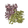
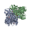
 Components Components | Glycine dehydrogenase [decarboxylating] | ||||||
 Keywords Keywords |  OXIDOREDUCTASE / alpha(2) homodimer / dehydrogenase (decarboxylating) / cofactor pyridoxal 5'-phosphate OXIDOREDUCTASE / alpha(2) homodimer / dehydrogenase (decarboxylating) / cofactor pyridoxal 5'-phosphate | ||||||
| Function / homology |  Function and homology information Function and homology informationglycine dehydrogenase (aminomethyl-transferring) / glycine dehydrogenase (decarboxylating) activity / glycine cleavage complex / glycine decarboxylation via glycine cleavage system / glycine binding / pyridoxal phosphate binding / cytosolSimilarity search - Function | ||||||
| Biological species |  | ||||||
| Method | X-RAY DIFFRACTION / SYNCHROTRON / MOLECULAR REPLACEMENT / Resolution: 1.899 Å | ||||||
 Authors Authors | Hasse, D. / Andersson, E. / Carlsson, G. / Masloboy, A. / Hagemann, M. / Bauwe, H. / Andersson, I. | ||||||
 Citation Citation | Journal: J.Biol.Chem. / Year: 2013 Title: Structure of the Homodimeric Glycine Decarboxylase P-protein from Synechocystis sp. PCC 6803 Suggests a Mechanism for Redox Regulation. Authors: Hasse, D. / Andersson, E. / Carlsson, G. / Masloboy, A. / Hagemann, M. / Bauwe, H. / Andersson, I. #1: Journal: Acta Crystallogr.,Sect.F / Year: 2010 Title: Crystallization and preliminary X-ray diffraction analyses of the homodimeric glycine decarboxylase (P-protein) from the cyanobacterium Synechocystis sp. PCC 6803. Authors: Hasse, D. / Hagemann, M. / Andersson, I. / Bauwe, H. | ||||||
| History |
|
- Structure visualization
| Structure viewer | Molecule: MolmilJmol/JSmol |
|---|
- Downloads & links
-Download
| PDBx/mmCIF format | 4lhc.cif.gz | 752.9 KB | Display | PDBx/mmCIF format |
|---|---|---|---|---|
| PDB format | pdb4lhc.ent.gz | 616.4 KB | Display | PDB format |
| PDBx/mmJSON format | 4lhc.json.gz | Tree view | PDBx/mmJSON format | |
| Others |  Other downloads Other downloads |
-Validation report
| Arichive directory | https://data.pdbj.org/pub/pdb/validation_reports/lh/4lhcftp://data.pdbj.org/pub/pdb/validation_reports/lh/4lhc | HTTPS FTP |
|---|
-Related structure data
| Related structure data |  4lglSC  4lhdC S: Starting model for refinement C: citing same article ( |
|---|---|
| Similar structure data |


-Links
 PDBj
PDBj


- Assembly
| Deposited unit | 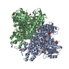
| ||||||||
|---|---|---|---|---|---|---|---|---|---|
| 1 |
| ||||||||
| Unit cell |
| ||||||||
| Components on special symmetry positions |
|
-Components
-Protein , 1 types, 2 molecules AB
| #1: Protein | Mass: 107649.172 Da / Num. of mol.: 2 Source method: isolated from a genetically manipulated source Source: (gene. exp.) Escherichia coli (E. coli) / Strain (production host): LMG194References: UniProt: P74416, glycine dehydrogenase (aminomethyl-transferring) |
|---|
-Non-polymers , 6 types, 1425 molecules 


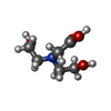

| #2: Chemical | ChemComp-GLY / Glycine Type: peptide linking / Mass: 75.067 Da / Num. of mol.: 5 / Source method: obtained synthetically / Formula: C2H5NO2 Type: peptide linking / Mass: 75.067 Da / Num. of mol.: 5 / Source method: obtained synthetically / Formula: C2H5NO2#3: Chemical | Bicarbonate Mass: 61.017 Da / Num. of mol.: 3 / Source method: obtained synthetically / Formula: CHO3 / Comment: pH buffer*YM Mass: 61.017 Da / Num. of mol.: 3 / Source method: obtained synthetically / Formula: CHO3 / Comment: pH buffer*YM#4: Chemical | ChemComp-PO4 / Phosphate Mass: 94.971 Da / Num. of mol.: 6 / Source method: obtained synthetically / Formula: PO4 Mass: 94.971 Da / Num. of mol.: 6 / Source method: obtained synthetically / Formula: PO4#5: Chemical | ChemComp-EDO / Ethylene glycol Mass: 62.068 Da / Num. of mol.: 16 / Source method: obtained synthetically / Formula: C2H6O2 Mass: 62.068 Da / Num. of mol.: 16 / Source method: obtained synthetically / Formula: C2H6O2#6: Chemical | ChemComp-BCN / | Bicine Mass: 163.172 Da / Num. of mol.: 1 / Source method: obtained synthetically / Formula: C6H13NO4 / Comment: pH buffer*YM Mass: 163.172 Da / Num. of mol.: 1 / Source method: obtained synthetically / Formula: C6H13NO4 / Comment: pH buffer*YM#7: Water | ChemComp-HOH / | WaterMass: 18.015 Da / Num. of mol.: 1394 / Source method: isolated from a natural source / Formula: H2O |
|---|
-Experimental details
-Experiment
| Experiment | Method: X-RAY DIFFRACTION / Number of used crystals: 1 |
|---|
- Sample preparation
| Crystal | Density Matthews: 2.74 Å3/Da / Density % sol: 55.13 % |
|---|---|
| Crystal grow | Temperature: 293 K / Method: vapor diffusion / pH: 5 Details: 100 mM Bicine pH 9, 1.6 M (NH4)3PO4, 5 mM TCEP and 0.2 mM PLP, VAPOR DIFFUSION, temperature 293K |
-Data collection
| Diffraction | Mean temperature: 100 K |
|---|---|
| Diffraction source | Source: SYNCHROTRON / Site: ESRF  / Beamline: ID14-1 / Wavelength: 0.9334 Å / Beamline: ID14-1 / Wavelength: 0.9334 Å |
| Detector | Type: ADSC QUANTUM 315r / Detector: CCD / Date: Apr 1, 2011 / Details: multilayer mirror |
| Radiation | Monochromator: Diamond (111), Ge(220) / Protocol: SINGLE WAVELENGTH / Monochromatic (M) / Laue (L): M / Scattering type: x-ray |
| Radiation wavelength | Wavelength: 0.9334 Å / Relative weight: 1 |
| Reflection | Resolution: 1.899→99 Å / Num. all: 185699 / Num. obs: 185454 / % possible obs: 99.9 % / Observed criterion σ(F): 0 / Observed criterion σ(I): 0 / Redundancy: 5.4 % / Rmerge(I) obs: 0.068 / Net I/σ(I): 18.4 |
| Reflection shell | Resolution: 1.899→1.97 Å / Redundancy: 4.4 % / Rmerge(I) obs: 0.371 / Mean I/σ(I) obs: 4.3 / % possible all: 99.1 |
- Processing
| Software |
| |||||||||||||||||||||||||||||||||||||||||||||||||||||||||||||||||||||||||||||||||||||||||||||||||||||||||||||||||||||||||||||||||||||||||||||||||||||||||||||||||||||||||||||||||||||||||||||||||||||||||||||||||||||||||
|---|---|---|---|---|---|---|---|---|---|---|---|---|---|---|---|---|---|---|---|---|---|---|---|---|---|---|---|---|---|---|---|---|---|---|---|---|---|---|---|---|---|---|---|---|---|---|---|---|---|---|---|---|---|---|---|---|---|---|---|---|---|---|---|---|---|---|---|---|---|---|---|---|---|---|---|---|---|---|---|---|---|---|---|---|---|---|---|---|---|---|---|---|---|---|---|---|---|---|---|---|---|---|---|---|---|---|---|---|---|---|---|---|---|---|---|---|---|---|---|---|---|---|---|---|---|---|---|---|---|---|---|---|---|---|---|---|---|---|---|---|---|---|---|---|---|---|---|---|---|---|---|---|---|---|---|---|---|---|---|---|---|---|---|---|---|---|---|---|---|---|---|---|---|---|---|---|---|---|---|---|---|---|---|---|---|---|---|---|---|---|---|---|---|---|---|---|---|---|---|---|---|---|---|---|---|---|---|---|---|---|---|---|---|---|---|---|---|---|
| Refinement | Method to determine structure: MOLECULAR REPLACEMENT Starting model: PDB ENTRY 4LGL Resolution: 1.899→49.783 Å / SU ML: 0.18 / σ(F): 1.34 / Phase error: 17.31 / Stereochemistry target values: ML
| |||||||||||||||||||||||||||||||||||||||||||||||||||||||||||||||||||||||||||||||||||||||||||||||||||||||||||||||||||||||||||||||||||||||||||||||||||||||||||||||||||||||||||||||||||||||||||||||||||||||||||||||||||||||||
| Solvent computation | Shrinkage radii: 1.11 Å / VDW probe radii: 1.3 Å / Solvent model: FLAT BULK SOLVENT MODEL / Bsol: 33.894 Å2 / ksol: 0.38 e/Å3 | |||||||||||||||||||||||||||||||||||||||||||||||||||||||||||||||||||||||||||||||||||||||||||||||||||||||||||||||||||||||||||||||||||||||||||||||||||||||||||||||||||||||||||||||||||||||||||||||||||||||||||||||||||||||||
| Displacement parameters |
| |||||||||||||||||||||||||||||||||||||||||||||||||||||||||||||||||||||||||||||||||||||||||||||||||||||||||||||||||||||||||||||||||||||||||||||||||||||||||||||||||||||||||||||||||||||||||||||||||||||||||||||||||||||||||
| Refinement step | Cycle: LAST / Resolution: 1.899→49.783 Å
| |||||||||||||||||||||||||||||||||||||||||||||||||||||||||||||||||||||||||||||||||||||||||||||||||||||||||||||||||||||||||||||||||||||||||||||||||||||||||||||||||||||||||||||||||||||||||||||||||||||||||||||||||||||||||
| Refine LS restraints |
| |||||||||||||||||||||||||||||||||||||||||||||||||||||||||||||||||||||||||||||||||||||||||||||||||||||||||||||||||||||||||||||||||||||||||||||||||||||||||||||||||||||||||||||||||||||||||||||||||||||||||||||||||||||||||
| LS refinement shell |
| |||||||||||||||||||||||||||||||||||||||||||||||||||||||||||||||||||||||||||||||||||||||||||||||||||||||||||||||||||||||||||||||||||||||||||||||||||||||||||||||||||||||||||||||||||||||||||||||||||||||||||||||||||||||||
| Refinement TLS params. | Method: refined / Refine-ID: X-RAY DIFFRACTION
| |||||||||||||||||||||||||||||||||||||||||||||||||||||||||||||||||||||||||||||||||||||||||||||||||||||||||||||||||||||||||||||||||||||||||||||||||||||||||||||||||||||||||||||||||||||||||||||||||||||||||||||||||||||||||
| Refinement TLS group |
|
