 Movie
Movie Controller

[English] 日本語
 Yorodumi
- PDB-4lgb: ABA-mimicking ligand N-(1-METHYL-2-OXO-1,2,3,4-TETRAHYDROQUINOLIN... -
+ Open data

- Basic information
| Entry | Database: PDB / ID: 4lgb | ||||||
|---|---|---|---|---|---|---|---|
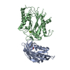
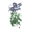
| Title | ABA-mimicking ligand N-(1-METHYL-2-OXO-1,2,3,4-TETRAHYDROQUINOLIN-6-YL)-1-(4-METHYLPHENYL)METHANESULFONAMIDE in complex with ABA receptor PYL2 and PP2C HAB1 | ||||||
 Components Components |
| ||||||
 Keywords Keywords | Hydrolase/receptor / abscisic acid mimicking compounds / PYR/PYL/RCAR / PP2C / Hydrolase-receptor complex | ||||||
| Function / homology |  Function and homology information Function and homology informationprotein phosphatase inhibitor complex /  abscisic acid binding / abscisic acid-activated signaling pathway / protein phosphatase inhibitor activity / myosin phosphatase activity / protein serine/threonine phosphatase activity / protein-serine/threonine phosphatase / signaling receptor activity / protein homodimerization activity / identical protein binding ...protein phosphatase inhibitor complex / abscisic acid binding / abscisic acid-activated signaling pathway / protein phosphatase inhibitor activity / myosin phosphatase activity / protein serine/threonine phosphatase activity / protein-serine/threonine phosphatase / signaling receptor activity / protein homodimerization activity / identical protein binding / metal ion binding / nucleus / plasma membrane / cytoplasm abscisic acid binding / abscisic acid-activated signaling pathway / protein phosphatase inhibitor activity / myosin phosphatase activity / protein serine/threonine phosphatase activity / protein-serine/threonine phosphatase / signaling receptor activity / protein homodimerization activity / identical protein binding ...protein phosphatase inhibitor complex / abscisic acid binding / abscisic acid-activated signaling pathway / protein phosphatase inhibitor activity / myosin phosphatase activity / protein serine/threonine phosphatase activity / protein-serine/threonine phosphatase / signaling receptor activity / protein homodimerization activity / identical protein binding / metal ion binding / nucleus / plasma membrane / cytoplasmSimilarity search - Function | ||||||
| Biological species |  Arabidopsis thaliana (thale cress) Arabidopsis thaliana (thale cress) | ||||||
| Method | X-RAY DIFFRACTION / SYNCHROTRON / MOLECULAR REPLACEMENT / Resolution: 3.15 Å | ||||||
 Authors Authors | Zhou, X.E. / Gao, M. / Liu, X. / Zhang, Y. / Xue, X. / Melcher, K. / Gao, P. / Wang, F. / Zeng, L. / Zhao, Y. ...Zhou, X.E. / Gao, M. / Liu, X. / Zhang, Y. / Xue, X. / Melcher, K. / Gao, P. / Wang, F. / Zeng, L. / Zhao, Y. / Zhao, Y. / Deng, P. / Zhong, D. / Zhu, J.-K. / Xu, Y. / Xu, H.E. | ||||||
 Citation Citation | Journal: Cell Res. / Year: 2013 Title: An ABA-mimicking ligand that reduces water loss and promotes drought resistance in plants. Authors: Cao, M. / Liu, X. / Zhang, Y. / Xue, X. / Zhou, X.E. / Melcher, K. / Gao, P. / Wang, F. / Zeng, L. / Zhao, Y. / Zhao, Y. / Deng, P. / Zhong, D. / Zhu, J.K. / Xu, H.E. / Xu, Y. | ||||||
| History |
|
- Structure visualization
| Structure viewer | Molecule: MolmilJmol/JSmol |
|---|
- Downloads & links
-Download
| PDBx/mmCIF format | 4lgb.cif.gz | 110.5 KB | Display | PDBx/mmCIF format |
|---|---|---|---|---|
| PDB format | pdb4lgb.ent.gz | 81.6 KB | Display | PDB format |
| PDBx/mmJSON format | 4lgb.json.gz | Tree view | PDBx/mmJSON format | |
| Others |  Other downloads Other downloads |
-Validation report
| Arichive directory | https://data.pdbj.org/pub/pdb/validation_reports/lg/4lgbftp://data.pdbj.org/pub/pdb/validation_reports/lg/4lgb | HTTPS FTP |
|---|
-Related structure data
| Related structure data |  4lg5C  4lgaC 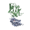 3kb3S C: citing same article ( S: Starting model for refinement |
|---|---|
| Similar structure data |


-Links
 PDBj
PDBj- Assembly
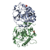
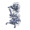
| Deposited unit | 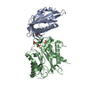
| ||||||||
|---|---|---|---|---|---|---|---|---|---|
| 1 |
| ||||||||
| Unit cell |
|
-Components
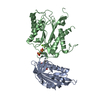
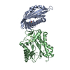
-Protein , 2 types, 2 molecules AB
| #1: Protein | Mass: 19905.414 Da / Num. of mol.: 1 / Fragment: unp residues 14-188 Source method: isolated from a genetically manipulated source Source: (gene. exp.) Arabidopsis thaliana (thale cress) / Gene: PYL2, RCAR14, At2g26040, T19L18.15 / Production host:  Escherichia coli (E. coli) / Strain (production host): BL21(DE3) / References: UniProt: O80992 Escherichia coli (E. coli) / Strain (production host): BL21(DE3) / References: UniProt: O80992 |
|---|---|
| #2: Protein | Mass: 37826.340 Da / Num. of mol.: 1 / Fragment: unp residues 172-511 Source method: isolated from a genetically manipulated source Source: (gene. exp.) Arabidopsis thaliana (thale cress) / Gene: At1g72770, F28P22.4, HAB1, P2C-HA / Plasmid: pETDuet / Production host: Escherichia coli (E. coli) / Strain (production host): BL21(DE3)References: UniProt: Q9CAJ0, protein-serine/threonine phosphatase |
-Non-polymers , 4 types, 38 molecules 


| #3: Chemical | ChemComp-1W7 /  Mass: 344.428 Da / Num. of mol.: 1 / Source method: obtained synthetically / Formula: C18H20N2O3S Mass: 344.428 Da / Num. of mol.: 1 / Source method: obtained synthetically / Formula: C18H20N2O3S | ||
|---|---|---|---|
| #4: Chemical | ChemComp-MG /  Mass: 24.305 Da / Num. of mol.: 1 / Source method: obtained synthetically / Formula: Mg Mass: 24.305 Da / Num. of mol.: 1 / Source method: obtained synthetically / Formula: Mg | ||
| #5: Chemical | Sulfate Mass: 96.063 Da / Num. of mol.: 3 / Source method: obtained synthetically / Formula: SO4 Mass: 96.063 Da / Num. of mol.: 3 / Source method: obtained synthetically / Formula: SO4#6: Water | ChemComp-HOH / | WaterMass: 18.015 Da / Num. of mol.: 33 / Source method: isolated from a natural source / Formula: H2O |
-Experimental details
-Experiment
| Experiment | Method: X-RAY DIFFRACTION / Number of used crystals: 1 |
|---|
- Sample preparation
| Crystal | Density Matthews: 2.52 Å3/Da / Density % sol: 51.09 % |
|---|---|
| Crystal grow | Temperature: 293 K / Method: vapor diffusion, hanging drop / pH: 6 Details: PEG3350, ammonium sulfate, pH 6.0, VAPOR DIFFUSION, HANGING DROP, temperature 293K |
-Data collection
| Diffraction | Mean temperature: 100 K | |||||||||||||||||||||||||||||||||||||||||||||||||||||||
|---|---|---|---|---|---|---|---|---|---|---|---|---|---|---|---|---|---|---|---|---|---|---|---|---|---|---|---|---|---|---|---|---|---|---|---|---|---|---|---|---|---|---|---|---|---|---|---|---|---|---|---|---|---|---|---|---|
| Diffraction source | Source: SYNCHROTRON / Site: SSRF  / Beamline: BL17U / Wavelength: 1 Å / Beamline: BL17U / Wavelength: 1 Å | |||||||||||||||||||||||||||||||||||||||||||||||||||||||
| Detector | Type: ADSC QUANTUM 315r / Detector: CCD / Date: Dec 18, 2012 | |||||||||||||||||||||||||||||||||||||||||||||||||||||||
| Radiation | Protocol: SINGLE WAVELENGTH / Monochromatic (M) / Laue (L): M / Scattering type: x-ray | |||||||||||||||||||||||||||||||||||||||||||||||||||||||
| Radiation wavelength | Wavelength: 1 Å / Relative weight: 1 | |||||||||||||||||||||||||||||||||||||||||||||||||||||||
| Reflection | Resolution: 3.15→30 Å / Num. all: 10654 / Num. obs: 8417 / % possible obs: 79 % / Observed criterion σ(F): 2 / Observed criterion σ(I): 2 / Redundancy: 4.4 % / Rmerge(I) obs: 0.168 / Net I/σ(I): 4.9 | |||||||||||||||||||||||||||||||||||||||||||||||||||||||
| Reflection shell |
|
- Processing
| Software |
| ||||||||||||||||||||||||||||||||||||||||||||||||||||||||||||||||||||||||||||||||||||||||||||||||||||||||||||||||||||||||||||||||||||||||||||||||||||||||||||||||||||||||||||||||||||||
|---|---|---|---|---|---|---|---|---|---|---|---|---|---|---|---|---|---|---|---|---|---|---|---|---|---|---|---|---|---|---|---|---|---|---|---|---|---|---|---|---|---|---|---|---|---|---|---|---|---|---|---|---|---|---|---|---|---|---|---|---|---|---|---|---|---|---|---|---|---|---|---|---|---|---|---|---|---|---|---|---|---|---|---|---|---|---|---|---|---|---|---|---|---|---|---|---|---|---|---|---|---|---|---|---|---|---|---|---|---|---|---|---|---|---|---|---|---|---|---|---|---|---|---|---|---|---|---|---|---|---|---|---|---|---|---|---|---|---|---|---|---|---|---|---|---|---|---|---|---|---|---|---|---|---|---|---|---|---|---|---|---|---|---|---|---|---|---|---|---|---|---|---|---|---|---|---|---|---|---|---|---|---|---|
| Refinement | Method to determine structure: MOLECULAR REPLACEMENT Starting model: pdb entry 3KB3 Resolution: 3.15→30 Å / Cor.coef. Fo:Fc: 0.882 / Cor.coef. Fo:Fc free: 0.864 / Occupancy max: 1 / Occupancy min: 1 / SU B: 32.772 / SU ML: 0.504 / Cross valid method: THROUGHOUT / ESU R Free: 0.73 / Stereochemistry target values: MAXIMUM LIKELIHOOD / Details: HYDROGENS HAVE BEEN ADDED IN THE RIDING POSITIONS
| ||||||||||||||||||||||||||||||||||||||||||||||||||||||||||||||||||||||||||||||||||||||||||||||||||||||||||||||||||||||||||||||||||||||||||||||||||||||||||||||||||||||||||||||||||||||
| Solvent computation | Ion probe radii: 0.8 Å / Shrinkage radii: 0.8 Å / VDW probe radii: 1.4 Å / Solvent model: BABINET MODEL WITH MASK | ||||||||||||||||||||||||||||||||||||||||||||||||||||||||||||||||||||||||||||||||||||||||||||||||||||||||||||||||||||||||||||||||||||||||||||||||||||||||||||||||||||||||||||||||||||||
| Displacement parameters | Biso mean: 57.048 Å2
| ||||||||||||||||||||||||||||||||||||||||||||||||||||||||||||||||||||||||||||||||||||||||||||||||||||||||||||||||||||||||||||||||||||||||||||||||||||||||||||||||||||||||||||||||||||||
| Refinement step | Cycle: LAST / Resolution: 3.15→30 Å
| ||||||||||||||||||||||||||||||||||||||||||||||||||||||||||||||||||||||||||||||||||||||||||||||||||||||||||||||||||||||||||||||||||||||||||||||||||||||||||||||||||||||||||||||||||||||
| Refine LS restraints |
|