 Movie
Movie Controller

[English] 日本語
 Yorodumi
- PDB-4lcm: Simvastatin Synthase (LOVD), from Aspergillus Terreus, LovD9 muta... -
+ Open data

- Basic information
| Entry | Database: PDB / ID: 4lcm | ||||||
|---|---|---|---|---|---|---|---|
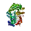
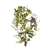
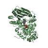
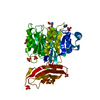
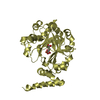
| Title | Simvastatin Synthase (LOVD), from Aspergillus Terreus, LovD9 mutant (simh9014) | ||||||
 Components Components | Transesterase | ||||||
 Keywords Keywords |  TRANSFERASE / laboratory-directed evolution / Transesterase TRANSFERASE / laboratory-directed evolution / Transesterase | ||||||
| Function / homology |  Function and homology information Function and homology informationmonacolin J acid methylbutanoate transferase / lovastatin biosynthetic process / polyketide synthase activity / polyketide biosynthetic process / acyltransferase activity / antibiotic biosynthetic process / defense response to fungus / hydrolase activitySimilarity search - Function | ||||||
| Biological species |  Aspergillus terreus (mold) Aspergillus terreus (mold) | ||||||
| Method | X-RAY DIFFRACTION / SYNCHROTRON / MOLECULAR REPLACEMENT / molecular replacement / Resolution: 3.19 Å | ||||||
 Authors Authors | Gao, X. / Sawaya, M.R. / Yeates, T.O. / Tang, Y. | ||||||
 Citation Citation | Journal: Nat.Chem.Biol. / Year: 2014 Title: The role of distant mutations and allosteric regulation on LovD active site dynamics. Authors: Jimenez-Oses, G. / Osuna, S. / Gao, X. / Sawaya, M.R. / Gilson, L. / Collier, S.J. / Huisman, G.W. / Yeates, T.O. / Tang, Y. / Houk, K.N. | ||||||
| History |
|
- Structure visualization
| Structure viewer | Molecule: MolmilJmol/JSmol |
|---|
- Downloads & links
-Download
| PDBx/mmCIF format | 4lcm.cif.gz | 620.4 KB | Display | PDBx/mmCIF format |
|---|---|---|---|---|
| PDB format | pdb4lcm.ent.gz | 519.6 KB | Display | PDB format |
| PDBx/mmJSON format | 4lcm.json.gz | Tree view | PDBx/mmJSON format | |
| Others |  Other downloads Other downloads |
-Validation report
| Arichive directory | https://data.pdbj.org/pub/pdb/validation_reports/lc/4lcmftp://data.pdbj.org/pub/pdb/validation_reports/lc/4lcm | HTTPS FTP |
|---|
-Related structure data
| Related structure data |  4lclC  3hlcS S: Starting model for refinement C: citing same article ( |
|---|---|
| Similar structure data |


-Links
 PDBj
PDBj
- Assembly
| Deposited unit | 
| ||||||||
|---|---|---|---|---|---|---|---|---|---|
| 1 | 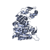
| ||||||||
| 2 | 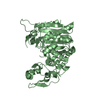
| ||||||||
| 3 | 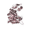
| ||||||||
| 4 | 
| ||||||||
| Unit cell |
| ||||||||
| Details | The biological unit is a monomer. There are four biological units in the asymmetric unit (chains A,B,C,D). |
-Components
| #1: Protein | Mass: 47271.008 Da / Num. of mol.: 4 Mutation: I4N A9V K26E R28S I35L C40A N43R C60N D96R S109C A123P M157V S164G S172N L174F A178V N191G L192I Q241M A247S R250K S256T A261H G275S Q297G L335M L361M V370I A383V N391S H404K Source method: isolated from a genetically manipulated source Source: (gene. exp.) Aspergillus terreus (mold) / Gene: lovD / Plasmid: pET28a / Production host:  Escherichia coli (E. coli) / Strain (production host): BL21 (DE3) / References: UniProt: Q9Y7D1 Escherichia coli (E. coli) / Strain (production host): BL21 (DE3) / References: UniProt: Q9Y7D1#2: Water | ChemComp-HOH / | Water Mass: 18.015 Da / Num. of mol.: 12 / Source method: isolated from a natural source / Formula: H2O Mass: 18.015 Da / Num. of mol.: 12 / Source method: isolated from a natural source / Formula: H2O |
|---|
-Experimental details
-Experiment
| Experiment | Method: X-RAY DIFFRACTION / Number of used crystals: 1 |
|---|
- Sample preparation
| Crystal | Density Matthews: 2.3 Å3/Da / Density % sol: 46.6 % |
|---|---|
| Crystal grow | Temperature: 298 K / Method: vapor diffusion, hanging drop / pH: 5.5 Details: 18% PEG-3350, 0.1 M Bis-Tris pH 5.5, 0.2 M MgCl2, 10mM DTT, VAPOR DIFFUSION, HANGING DROP, temperature 298K |
-Data collection
| Diffraction | Mean temperature: 100 K | |||||||||||||||||||||||||||||||||||||||||||||||||||||||||||||||||||||||||||||||||||||||||||||||||||||||||||||||||||||||||||||||||||||||||||||||||||
|---|---|---|---|---|---|---|---|---|---|---|---|---|---|---|---|---|---|---|---|---|---|---|---|---|---|---|---|---|---|---|---|---|---|---|---|---|---|---|---|---|---|---|---|---|---|---|---|---|---|---|---|---|---|---|---|---|---|---|---|---|---|---|---|---|---|---|---|---|---|---|---|---|---|---|---|---|---|---|---|---|---|---|---|---|---|---|---|---|---|---|---|---|---|---|---|---|---|---|---|---|---|---|---|---|---|---|---|---|---|---|---|---|---|---|---|---|---|---|---|---|---|---|---|---|---|---|---|---|---|---|---|---|---|---|---|---|---|---|---|---|---|---|---|---|---|---|---|---|
| Diffraction source | Source: SYNCHROTRON / Site: APS  / Beamline: 24-ID-E / Wavelength: 0.9791 Å / Beamline: 24-ID-E / Wavelength: 0.9791 Å | |||||||||||||||||||||||||||||||||||||||||||||||||||||||||||||||||||||||||||||||||||||||||||||||||||||||||||||||||||||||||||||||||||||||||||||||||||
| Detector | Type: ADSC QUANTUM 315 / Detector: CCD / Date: Jul 22, 2011 | |||||||||||||||||||||||||||||||||||||||||||||||||||||||||||||||||||||||||||||||||||||||||||||||||||||||||||||||||||||||||||||||||||||||||||||||||||
| Radiation | Protocol: SINGLE WAVELENGTH / Monochromatic (M) / Laue (L): M / Scattering type: x-ray | |||||||||||||||||||||||||||||||||||||||||||||||||||||||||||||||||||||||||||||||||||||||||||||||||||||||||||||||||||||||||||||||||||||||||||||||||||
| Radiation wavelength | Wavelength: 0.9791 Å / Relative weight: 1 | |||||||||||||||||||||||||||||||||||||||||||||||||||||||||||||||||||||||||||||||||||||||||||||||||||||||||||||||||||||||||||||||||||||||||||||||||||
| Reflection | Resolution: 3.19→19.66 Å / Num. all: 25955 / Num. obs: 25955 / % possible obs: 92.1 % / Observed criterion σ(I): -3 / Redundancy: 1.8 % / Biso Wilson estimate: 37.052 Å2 / Rmerge(I) obs: 0.175 / Net I/σ(I): 4.6 | |||||||||||||||||||||||||||||||||||||||||||||||||||||||||||||||||||||||||||||||||||||||||||||||||||||||||||||||||||||||||||||||||||||||||||||||||||
| Reflection shell | Diffraction-ID: 1
|
-Phasing
| Phasing | Method: molecular replacement | |||||||||
|---|---|---|---|---|---|---|---|---|---|---|
| Phasing MR | Model details: Phaser MODE: MR_AUTO
|
- Processing
| Software |
| |||||||||||||||||||||||||||||||||||||||||||||||||||||||||||||||||||||||||||||||||||||||||||||||||||||||||||||||||||||||||||||
|---|---|---|---|---|---|---|---|---|---|---|---|---|---|---|---|---|---|---|---|---|---|---|---|---|---|---|---|---|---|---|---|---|---|---|---|---|---|---|---|---|---|---|---|---|---|---|---|---|---|---|---|---|---|---|---|---|---|---|---|---|---|---|---|---|---|---|---|---|---|---|---|---|---|---|---|---|---|---|---|---|---|---|---|---|---|---|---|---|---|---|---|---|---|---|---|---|---|---|---|---|---|---|---|---|---|---|---|---|---|---|---|---|---|---|---|---|---|---|---|---|---|---|---|---|---|---|
| Refinement | Method to determine structure: MOLECULAR REPLACEMENT Starting model: 3HLC Resolution: 3.19→19.66 Å / Cor.coef. Fo:Fc: 0.8573 / Cor.coef. Fo:Fc free: 0.7961 / Occupancy max: 1 / Occupancy min: 1 / Cross valid method: THROUGHOUT / σ(F): 0 / Stereochemistry target values: Engh & Huber
| |||||||||||||||||||||||||||||||||||||||||||||||||||||||||||||||||||||||||||||||||||||||||||||||||||||||||||||||||||||||||||||
| Displacement parameters | Biso max: 148.28 Å2 / Biso mean: 50.781 Å2 / Biso min: 3 Å2
| |||||||||||||||||||||||||||||||||||||||||||||||||||||||||||||||||||||||||||||||||||||||||||||||||||||||||||||||||||||||||||||
| Refine analyze | Luzzati coordinate error obs: 0.566 Å | |||||||||||||||||||||||||||||||||||||||||||||||||||||||||||||||||||||||||||||||||||||||||||||||||||||||||||||||||||||||||||||
| Refinement step | Cycle: LAST / Resolution: 3.19→19.66 Å
| |||||||||||||||||||||||||||||||||||||||||||||||||||||||||||||||||||||||||||||||||||||||||||||||||||||||||||||||||||||||||||||
| Refine LS restraints |
| |||||||||||||||||||||||||||||||||||||||||||||||||||||||||||||||||||||||||||||||||||||||||||||||||||||||||||||||||||||||||||||
| LS refinement shell | Resolution: 3.19→3.32 Å / Total num. of bins used: 13
| |||||||||||||||||||||||||||||||||||||||||||||||||||||||||||||||||||||||||||||||||||||||||||||||||||||||||||||||||||||||||||||
| Refinement TLS params. | Method: refined / Refine-ID: X-RAY DIFFRACTION
| |||||||||||||||||||||||||||||||||||||||||||||||||||||||||||||||||||||||||||||||||||||||||||||||||||||||||||||||||||||||||||||
| Refinement TLS group |
|
