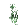
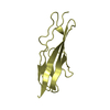
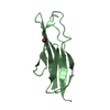
[English] 日本語
Yorodumi
- PDB-4l9d: Crystal structure of the PKD1 domain from Vibrio cholerae metallo... -

+
Open data


ID or keywords:

Loading...

-
Basic information

Entry
Database: PDB / ID: 4l9d
TitleCrystal structure of the PKD1 domain from Vibrio cholerae metalloprotease PrtV
ComponentsProtease
KeywordsCELL ADHESION / PKD domain
Function / homology
Function and homology information


cytolysis / Hydrolases; Acting on peptide bonds (peptidases); Metalloendopeptidases / : / metallopeptidase activity / extracellular region / metal ion binding
Similarity search - Function
Peptidase M6, InhA / Peptidase M6-like, domain / Immune inhibitor A peptidase M6, catalytic domain / PKD domain / PKD domain / Polycystic kidney disease (PKD) domain profile. / PKD domain / PKD domain superfamily / PKD/Chitinase domain / Repeats in polycystic kidney disease 1 (PKD1) and other proteins ...Peptidase M6, InhA / Peptidase M6-like, domain / Immune inhibitor A peptidase M6, catalytic domain / PKD domain / PKD domain / Polycystic kidney disease (PKD) domain profile. / PKD domain / PKD domain superfamily / PKD/Chitinase domain / Repeats in polycystic kidney disease 1 (PKD1) and other proteins / Immunoglobulins / Immunoglobulin-like fold / Immunoglobulin-like / Sandwich / Mainly Beta
Similarity search - Domain/homology
DI(HYDROXYETHYL)ETHER / Pre-pro-metalloprotease PrtV
Similarity search - Component
Biological speciesVibrio cholerae (bacteria)
MethodX-RAY DIFFRACTION / SYNCHROTRON / MOLECULAR REPLACEMENT / Resolution: 1.1 Å
AuthorsEdwin, A. / Stier, G. / Sauer-Eriksson, A.E.
CitationJournal: FEBS Open Bio / Year: 2013
Title: Calcium binding by the PKD1 domain regulates interdomain flexibility in Vibrio cholerae metalloprotease PrtV.
Authors: Edwin, A. / Rompikuntal, P. / Bjorn, E. / Stier, G. / Wai, S.N. / Sauer-Eriksson, A.E.
History
DepositionJun 18, 2013Deposition site: RCSB / Processing site: RCSB
Revision 1.0Jul 10, 2013Provider: repository / Type: Initial release
Revision 1.1Aug 14, 2013Group: Database references
Revision 1.2Feb 28, 2024Group: Data collection / Database references / Derived calculations
Category: chem_comp_atom / chem_comp_bond ...chem_comp_atom / chem_comp_bond / database_2 / pdbx_struct_conn_angle / struct_conn / struct_ref_seq_dif / struct_site
Item: _database_2.pdbx_DOI / _database_2.pdbx_database_accession ..._database_2.pdbx_DOI / _database_2.pdbx_database_accession / _pdbx_struct_conn_angle.ptnr1_auth_asym_id / _pdbx_struct_conn_angle.ptnr1_auth_comp_id / _pdbx_struct_conn_angle.ptnr1_auth_seq_id / _pdbx_struct_conn_angle.ptnr1_label_alt_id / _pdbx_struct_conn_angle.ptnr1_label_asym_id / _pdbx_struct_conn_angle.ptnr1_label_atom_id / _pdbx_struct_conn_angle.ptnr1_label_comp_id / _pdbx_struct_conn_angle.ptnr1_label_seq_id / _pdbx_struct_conn_angle.ptnr2_auth_asym_id / _pdbx_struct_conn_angle.ptnr2_auth_comp_id / _pdbx_struct_conn_angle.ptnr2_auth_seq_id / _pdbx_struct_conn_angle.ptnr2_label_asym_id / _pdbx_struct_conn_angle.ptnr2_label_atom_id / _pdbx_struct_conn_angle.ptnr2_label_comp_id / _pdbx_struct_conn_angle.ptnr3_auth_asym_id / _pdbx_struct_conn_angle.ptnr3_auth_comp_id / _pdbx_struct_conn_angle.ptnr3_auth_seq_id / _pdbx_struct_conn_angle.ptnr3_label_alt_id / _pdbx_struct_conn_angle.ptnr3_label_asym_id / _pdbx_struct_conn_angle.ptnr3_label_atom_id / _pdbx_struct_conn_angle.ptnr3_label_comp_id / _pdbx_struct_conn_angle.ptnr3_label_seq_id / _pdbx_struct_conn_angle.value / _struct_conn.pdbx_dist_value / _struct_conn.pdbx_ptnr1_label_alt_id / _struct_conn.ptnr1_auth_asym_id / _struct_conn.ptnr1_auth_comp_id / _struct_conn.ptnr1_auth_seq_id / _struct_conn.ptnr1_label_asym_id / _struct_conn.ptnr1_label_atom_id / _struct_conn.ptnr1_label_comp_id / _struct_conn.ptnr1_label_seq_id / _struct_conn.ptnr2_auth_asym_id / _struct_conn.ptnr2_auth_comp_id / _struct_conn.ptnr2_auth_seq_id / _struct_conn.ptnr2_label_asym_id / _struct_conn.ptnr2_label_atom_id / _struct_conn.ptnr2_label_comp_id / _struct_ref_seq_dif.details / _struct_site.pdbx_auth_asym_id / _struct_site.pdbx_auth_comp_id / _struct_site.pdbx_auth_seq_id

-
Structure visualization

Structure viewerMolecule:
MolmilJmol/JSmol

Downloads & links

-
Assembly

Deposited unit
A: Protease
B: Protease
hetero molecules


Theoretical massNumber of molelcules
Total (without water)19,20610
Polymers18,8842
Non-polymers3228
Water5,405300
1
A: Protease
hetero molecules


Theoretical massNumber of molelcules
Total (without water)9,5364
Polymers9,4421
Non-polymers943
Water181
TypeNameSymmetry operationNumber
identity operation1_555x,y,z1
MethodPISA
2
B: Protease
hetero molecules


Theoretical massNumber of molelcules
Total (without water)9,6706
Polymers9,4421
Non-polymers2285
Water181
TypeNameSymmetry operationNumber
identity operation1_555x,y,z1
MethodPISA
Unit cell
Length a, b, c (Å)41.800, 50.833, 67.297
Angle α, β, γ (deg.)90.00, 90.00, 90.00
Int Tables number19
Space group name H-MP212121

-
Components

-
Protein , 1 types, 2 molecules AB

#1: Protein Protease /


Mass: 9442.131 Da / Num. of mol.: 2 / Fragment: PKD1, UNP residues 755-839
Source method: isolated from a genetically manipulated source
Source: (gene. exp.) Vibrio cholerae (bacteria) / Strain: M66-2 / Gene: prtV, VCM66_A0219 / Production host: Escherichia coli (E. coli) / References: UniProt: C3LUP3

-
Non-polymers , 5 types, 308 molecules

#2: Chemical ChemComp-NA / SODIUM ION


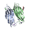
Mass: 22.990 Da / Num. of mol.: 3 / Source method: obtained synthetically / Formula: Na
#3: Chemical ChemComp-CL / CHLORIDE ION / Chloride


Mass: 35.453 Da / Num. of mol.: 3 / Source method: obtained synthetically / Formula: Cl
#4: Chemical ChemComp-CA / CALCIUM ION


Mass: 40.078 Da / Num. of mol.: 1 / Source method: obtained synthetically / Formula: Ca
#5: Chemical ChemComp-PEG / DI(HYDROXYETHYL)ETHER / Diethylene glycol


Mass: 106.120 Da / Num. of mol.: 1 / Source method: obtained synthetically / Formula: C4H10O3
#6: Water ChemComp-HOH / water / Water


Mass: 18.015 Da / Num. of mol.: 300 / Source method: isolated from a natural source / Formula: H2O

-
Experimental details

-
Experiment

ExperimentMethod: X-RAY DIFFRACTION / Number of used crystals: 1

-
Sample preparation

CrystalDensity Matthews: 1.89 Å3/Da / Density % sol: 35.02 %
Crystal growTemperature: 291 K / Method: vapor diffusion, hanging drop / pH: 5.5
Details: 1.2 M sodium citrate and 20% (w/v) PEG 8K, pH 5.5, VAPOR DIFFUSION, HANGING DROP, temperature 291K

-
Data collection

DiffractionMean temperature: 100 K
Diffraction sourceSource: SYNCHROTRON / Site: ESRF / Beamline: ID23-1 / Wavelength: 1.0398 Å
DetectorType: ADSC QUANTUM 315r / Detector: CCD / Date: May 29, 2011
RadiationMonochromator: Si 111 CHANNEL / Protocol: SINGLE WAVELENGTH / Monochromatic (M) / Laue (L): M / Scattering type: x-ray
Radiation wavelengthWavelength: 1.0398 Å / Relative weight: 1
ReflectionResolution: 1.1→12 Å / Num. obs: 58243 / % possible obs: 99 % / Observed criterion σ(F): 1 / Observed criterion σ(I): 1 / Rmerge(I) obs: 0.06 / Net I/σ(I): 16.7
Reflection shellResolution: 1.1→1.15 Å / % possible all: 96.2

-
Processing

Software
NameVersionClassification
XDSdata scaling
Auto-Rickshawphasing
REFMAC5.7.0032refinement
XDSdata reduction
SCALAdata scaling
RefinementMethod to determine structure: MOLECULAR REPLACEMENT / Resolution: 1.1→12 Å / Cor.coef. Fo:Fc: 0.98 / Cor.coef. Fo:Fc free: 0.97 / SU B: 0.77 / SU ML: 0.017 / Cross valid method: THROUGHOUT / ESU R: 0.026 / ESU R Free: 0.028 / Stereochemistry target values: MAXIMUM LIKELIHOOD / Details: HYDROGENS HAVE BEEN ADDED IN THE RIDING POSITIONS
RfactorNum. reflection% reflectionSelection details
Rfree0.1404 2958 5.1 %RANDOM
Rwork0.10947 ---
obs0.11099 55335 98.95 %-
Solvent computationIon probe radii: 0.8 Å / Shrinkage radii: 0.8 Å / VDW probe radii: 1.2 Å / Solvent model: MASK
Displacement parametersBiso mean: 10.249 Å2
Baniso -1Baniso -2Baniso -3
1--0.12 Å2-0 Å20 Å2
2--0.14 Å20 Å2
3----0.02 Å2
Refinement stepCycle: LAST / Resolution: 1.1→12 Å
ProteinNucleic acidLigandSolventTotal
Num. atoms1233 0 14 300 1547
Refine LS restraints
Refine-IDTypeDev idealDev ideal targetNumber
X-RAY DIFFRACTIONr_bond_refined_d0.020.021484
X-RAY DIFFRACTIONr_bond_other_d0.0040.021330
X-RAY DIFFRACTIONr_angle_refined_deg2.1921.9342047
X-RAY DIFFRACTIONr_angle_other_deg1.01233116
X-RAY DIFFRACTIONr_dihedral_angle_1_deg13.445211
X-RAY DIFFRACTIONr_dihedral_angle_2_deg36.46126.87564
X-RAY DIFFRACTIONr_dihedral_angle_3_deg11.92615250
X-RAY DIFFRACTIONr_dihedral_angle_4_deg10.466152
X-RAY DIFFRACTIONr_chiral_restr0.1290.2231
X-RAY DIFFRACTIONr_gen_planes_refined0.0120.021798
X-RAY DIFFRACTIONr_gen_planes_other0.0040.02324
X-RAY DIFFRACTIONr_nbd_refined
X-RAY DIFFRACTIONr_nbd_other
X-RAY DIFFRACTIONr_nbtor_refined
X-RAY DIFFRACTIONr_nbtor_other
X-RAY DIFFRACTIONr_xyhbond_nbd_refined
X-RAY DIFFRACTIONr_xyhbond_nbd_other
X-RAY DIFFRACTIONr_metal_ion_refined
X-RAY DIFFRACTIONr_metal_ion_other
X-RAY DIFFRACTIONr_symmetry_vdw_refined
X-RAY DIFFRACTIONr_symmetry_vdw_other
X-RAY DIFFRACTIONr_symmetry_hbond_refined
X-RAY DIFFRACTIONr_symmetry_hbond_other
X-RAY DIFFRACTIONr_symmetry_metal_ion_refined
X-RAY DIFFRACTIONr_symmetry_metal_ion_other
X-RAY DIFFRACTIONr_mcbond_it2.7370.677763
X-RAY DIFFRACTIONr_mcbond_other2.4680.667762
X-RAY DIFFRACTIONr_mcangle_it2.0241.007980
X-RAY DIFFRACTIONr_mcangle_other2.111.014981
X-RAY DIFFRACTIONr_scbond_it3.3260.944721
X-RAY DIFFRACTIONr_scbond_other3.3270.943721
X-RAY DIFFRACTIONr_scangle_it
X-RAY DIFFRACTIONr_scangle_other4.0851.3161061
X-RAY DIFFRACTIONr_long_range_B_refined4.7898.3021947
X-RAY DIFFRACTIONr_long_range_B_other4.0957.1051786
X-RAY DIFFRACTIONr_rigid_bond_restr6.14232813
X-RAY DIFFRACTIONr_sphericity_free36.289551
X-RAY DIFFRACTIONr_sphericity_bonded11.81953025
LS refinement shellResolution: 1.1→1.128 Å / Total num. of bins used: 20
RfactorNum. reflection% reflection
Rfree0.176 228 -
Rwork0.138 3837 -
obs--95.67 %

+
About Yorodumi

-
News

-
Feb 9, 2022. New format data for meta-information of EMDB entries

New format data for meta-information of EMDB entries

  • Version 3 of the EMDB header file is now the official format.
  • The previous official version 1.9 will be removed from the archive.

Related info.:EMDB header

External links:wwPDB to switch to version 3 of the EMDB data model

-
Aug 12, 2020. Covid-19 info

Covid-19 info

URL: https://pdbj.org/emnavi/covid19.php

New page: Covid-19 featured information page in EM Navigator.

Related info.:Covid-19 info / Mar 5, 2020. Novel coronavirus structure data

+
Mar 5, 2020. Novel coronavirus structure data

Novel coronavirus structure data

Related info.:Yorodumi Speices / Aug 12, 2020. Covid-19 info

External links:COVID-19 featured content - PDBj / Molecule of the Month (242):Coronavirus Proteases

+
Jan 31, 2019. EMDB accession codes are about to change! (news from PDBe EMDB page)

EMDB accession codes are about to change! (news from PDBe EMDB page)

  • The allocation of 4 digits for EMDB accession codes will soon come to an end. Whilst these codes will remain in use, new EMDB accession codes will include an additional digit and will expand incrementally as the available range of codes is exhausted. The current 4-digit format prefixed with “EMD-” (i.e. EMD-XXXX) will advance to a 5-digit format (i.e. EMD-XXXXX), and so on. It is currently estimated that the 4-digit codes will be depleted around Spring 2019, at which point the 5-digit format will come into force.
  • The EM Navigator/Yorodumi systems omit the EMD- prefix.

Related info.:Q: What is EMD? / ID/Accession-code notation in Yorodumi/EM Navigator

External links:EMDB Accession Codes are Changing Soon! / Contact to PDBj

+
Jul 12, 2017. Major update of PDB

Major update of PDB

  • wwPDB released updated PDB data conforming to the new PDBx/mmCIF dictionary.
  • This is a major update changing the version number from 4 to 5, and with Remediation, in which all the entries are updated.
  • In this update, many items about electron microscopy experimental information are reorganized (e.g. em_software).
  • Now, EM Navigator and Yorodumi are based on the updated data.

External links:wwPDB Remediation / Enriched Model Files Conforming to OneDep Data Standards Now Available in the PDB FTP Archive

-
Yorodumi

Thousand views of thousand structures

  • Yorodumi is a browser for structure data from EMDB, PDB, SASBDB, etc.
  • This page is also the successor to EM Navigator detail page, and also detail information page/front-end page for Omokage search.
  • The word "yorodu" (or yorozu) is an old Japanese word meaning "ten thousand". "mi" (miru) is to see.

Related info.:EMDB / PDB / SASBDB / Comparison of 3 databanks / Yorodumi Search / Aug 31, 2016. New EM Navigator & Yorodumi / Yorodumi Papers / Jmol/JSmol / Function and homology information / Changes in new EM Navigator and Yorodumi

Read more