 Movie
Movie Controller

[English] 日本語
 Yorodumi
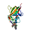
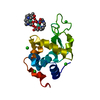
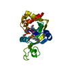
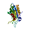
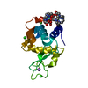
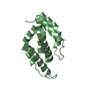
- PDB-4l9c: Crystal structure of the FP domain of human F-box protein Fbxo7 (... -
+ Open data

- Basic information
| Entry | Database: PDB / ID: 4l9c | ||||||
|---|---|---|---|---|---|---|---|
| Title | Crystal structure of the FP domain of human F-box protein Fbxo7 (native) | ||||||
 Components Components | F-box only protein 7 | ||||||
 Keywords Keywords |  PROTEIN BINDING / alpha/beta fold PROTEIN BINDING / alpha/beta fold | ||||||
| Function / homology |  Function and homology information Function and homology informationnegative regulation of lymphocyte differentiation / glial cytoplasmic inclusion / classical Lewy body / Lewy neurite / Lewy body corona / Lewy body core / positive regulation of autophagy of mitochondrion / lymphocyte differentiation / autophagy of mitochondrion / negative regulation of oxidative stress-induced neuron intrinsic apoptotic signaling pathway ...negative regulation of lymphocyte differentiation / glial cytoplasmic inclusion / classical Lewy body / Lewy neurite / Lewy body corona / Lewy body core / positive regulation of autophagy of mitochondrion / lymphocyte differentiation / autophagy of mitochondrion / negative regulation of oxidative stress-induced neuron intrinsic apoptotic signaling pathway / protein targeting to mitochondrion / regulation of locomotion / SCF ubiquitin ligase complex / negative regulation of G1/S transition of mitotic cell cycle / regulation of neuron projection development / ubiquitin-like ligase-substrate adaptor activity / protein K48-linked ubiquitination / ubiquitin ligase complex / ubiquitin binding / regulation of protein stability / ubiquitin-protein transferase activity / Antigen processing: Ubiquitination & Proteasome degradation / Neddylation / ubiquitin-dependent protein catabolic process / proteasome-mediated ubiquitin-dependent protein catabolic process / protein ubiquitination / protein heterodimerization activity / ubiquitin protein ligase binding / protein kinase binding / protein-containing complex / mitochondrion / nucleoplasm / nucleus / cytosol / cytoplasmSimilarity search - Function | ||||||
| Biological species |  Homo sapiens (human) Homo sapiens (human) | ||||||
| Method | X-RAY DIFFRACTION / SYNCHROTRON / MOLECULAR REPLACEMENT / Resolution: 2.1 Å | ||||||
 Authors Authors | Du, Z. / Huang, X. / Shang, J. / Yang, Y. / Wang, G. | ||||||
 Citation Citation | Journal: Acta Crystallogr.,Sect.D / Year: 2014 Title: Structure of the FP domain of Fbxo7 reveals a novel mode of protein-protein interaction. Authors: Shang, J. / Wang, G. / Yang, Y. / Huang, X. / Du, Z. | ||||||
| History |
|
- Structure visualization
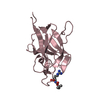
| Structure viewer | Molecule: MolmilJmol/JSmol |
|---|
- Downloads & links
-Download
| PDBx/mmCIF format | 4l9c.cif.gz | 73 KB | Display | PDBx/mmCIF format |
|---|---|---|---|---|
| PDB format | pdb4l9c.ent.gz | 54.3 KB | Display | PDB format |
| PDBx/mmJSON format | 4l9c.json.gz | Tree view | PDBx/mmJSON format | |
| Others |  Other downloads Other downloads |
-Validation report
| Arichive directory | https://data.pdbj.org/pub/pdb/validation_reports/l9/4l9cftp://data.pdbj.org/pub/pdb/validation_reports/l9/4l9c | HTTPS FTP |
|---|
-Related structure data


-Links
 PDBj
PDBj


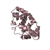
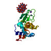
- Assembly
| Deposited unit | 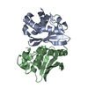
| ||||||||
|---|---|---|---|---|---|---|---|---|---|
| 1 | 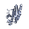
| ||||||||
| 2 | 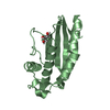
| ||||||||
| 3 | 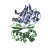
| ||||||||
| Unit cell |
|
-Components
| #1: Protein | Mass: 17761.744 Da / Num. of mol.: 2 / Fragment: unp residues 180-335 Source method: isolated from a genetically manipulated source Source: (gene. exp.) Homo sapiens (human) / Gene: FBX7, FBXO7, HUMAN FBXO7 / Production host:  Escherichia coli (E. coli) / Strain (production host): BL21(DE3) / References: UniProt: Q9Y3I1 Escherichia coli (E. coli) / Strain (production host): BL21(DE3) / References: UniProt: Q9Y3I1#2: Chemical | ChemComp-GOL / | Glycerol  Mass: 92.094 Da / Num. of mol.: 1 / Source method: obtained synthetically / Formula: C3H8O3 Mass: 92.094 Da / Num. of mol.: 1 / Source method: obtained synthetically / Formula: C3H8O3#3: Water | ChemComp-HOH / | Water Mass: 18.015 Da / Num. of mol.: 96 / Source method: isolated from a natural source / Formula: H2O Mass: 18.015 Da / Num. of mol.: 96 / Source method: isolated from a natural source / Formula: H2O |
|---|
-Experimental details
-Experiment
| Experiment | Method: X-RAY DIFFRACTION / Number of used crystals: 1 |
|---|
- Sample preparation
| Crystal | Density Matthews: 1.79 Å3/Da / Density % sol: 31.29 % |
|---|---|
| Crystal grow | Temperature: 298 K / Method: vapor diffusion, hanging drop / pH: 8 Details: PEG 3350, MES 7.2, KBr , pH 8.0, VAPOR DIFFUSION, HANGING DROP, temperature 298K |
-Data collection
| Diffraction | Mean temperature: 100 K |
|---|---|
| Diffraction source | Source: SYNCHROTRON / Site: APS  / Beamline: 21-ID-D / Wavelength: 0.97872 Å / Beamline: 21-ID-D / Wavelength: 0.97872 Å |
| Detector | Type: MARMOSAIC 225 mm CCD / Detector: CCD |
| Radiation | Protocol: SINGLE WAVELENGTH / Monochromatic (M) / Laue (L): M / Scattering type: x-ray |
| Radiation wavelength | Wavelength: 0.97872 Å / Relative weight: 1 |
| Reflection | Highest resolution: 2.1 Å / Num. obs: 15476 |
- Processing
| Software |
| ||||||||||||||||||||||||||||||||||||||||||||||||||||||||||||||||||||||||||||||||||||
|---|---|---|---|---|---|---|---|---|---|---|---|---|---|---|---|---|---|---|---|---|---|---|---|---|---|---|---|---|---|---|---|---|---|---|---|---|---|---|---|---|---|---|---|---|---|---|---|---|---|---|---|---|---|---|---|---|---|---|---|---|---|---|---|---|---|---|---|---|---|---|---|---|---|---|---|---|---|---|---|---|---|---|---|---|---|
| Refinement | Method to determine structure: MOLECULAR REPLACEMENT / Resolution: 2.1→39.628 Å / Occupancy max: 1 / Occupancy min: 1 / FOM work R set: 0.8234 / SU ML: 0.24 / σ(F): 1.35 / Phase error: 23.99 / Stereochemistry target values: ML
| ||||||||||||||||||||||||||||||||||||||||||||||||||||||||||||||||||||||||||||||||||||
| Solvent computation | Shrinkage radii: 0.9 Å / VDW probe radii: 1.11 Å / Solvent model: FLAT BULK SOLVENT MODEL | ||||||||||||||||||||||||||||||||||||||||||||||||||||||||||||||||||||||||||||||||||||
| Displacement parameters | Biso max: 90.45 Å2 / Biso mean: 32.2989 Å2 / Biso min: 6.46 Å2 | ||||||||||||||||||||||||||||||||||||||||||||||||||||||||||||||||||||||||||||||||||||
| Refinement step | Cycle: LAST / Resolution: 2.1→39.628 Å
| ||||||||||||||||||||||||||||||||||||||||||||||||||||||||||||||||||||||||||||||||||||
| Refine LS restraints |
| ||||||||||||||||||||||||||||||||||||||||||||||||||||||||||||||||||||||||||||||||||||
| LS refinement shell | Refine-ID: X-RAY DIFFRACTION / Total num. of bins used: 11
|
