 Movie
Movie Controller

[English] 日本語
 Yorodumi
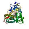
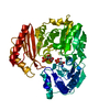
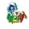
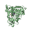
- PDB-4l7p: Human artd3 (parp3) - catalytic domain in complex with inhibitor ... -
+ Open data

- Basic information
| Entry | Database: PDB / ID: 4l7p | ||||||
|---|---|---|---|---|---|---|---|
| Title | Human artd3 (parp3) - catalytic domain in complex with inhibitor ME0395 | ||||||
 Components Components | Poly [ADP-ribose] polymerase 3 | ||||||
 Keywords Keywords | transferase/transferase inhibitor / DIPHTHERIA TOXIN LIKE ADP-RIBOSE TRASNFERASE /  TRANSFERASE / ADP-RIBOSYLATION / transferase-transferase inhibitor complex TRANSFERASE / ADP-RIBOSYLATION / transferase-transferase inhibitor complex | ||||||
| Function / homology |  Function and homology information Function and homology informationnegative regulation of isotype switching / negative regulation of telomerase RNA reverse transcriptase activity / NAD+- protein-lysine ADP-ribosyltransferase activity / positive regulation of DNA ligation / NAD DNA ADP-ribosyltransferase activity / NAD+- protein-aspartate ADP-ribosyltransferase activity / NAD+-protein-glutamate ADP-ribosyltransferase activity / DNA ADP-ribosylation / protein localization to site of double-strand break / protein auto-ADP-ribosylation ...negative regulation of isotype switching / negative regulation of telomerase RNA reverse transcriptase activity / NAD+- protein-lysine ADP-ribosyltransferase activity / positive regulation of DNA ligation / NAD DNA ADP-ribosyltransferase activity / NAD+- protein-aspartate ADP-ribosyltransferase activity / NAD+-protein-glutamate ADP-ribosyltransferase activity / DNA ADP-ribosylation / protein localization to site of double-strand break / protein auto-ADP-ribosylation / intercellular bridge / positive regulation of double-strand break repair via nonhomologous end joining / NAD+-protein ADP-ribosyltransferase activity / NAD+ ADP-ribosyltransferase activity / Transferases; Glycosyltransferases; Pentosyltransferases / catalytic activity / regulation of mitotic spindle organization / centriole / telomere maintenance / nucleotidyltransferase activity / double-strand break repair / site of double-strand break / nuclear body / centrosome / nucleolus / nucleoplasm / cytoplasmSimilarity search - Function | ||||||
| Biological species |  Homo sapiens (human) Homo sapiens (human) | ||||||
| Method | X-RAY DIFFRACTION / SYNCHROTRON / MOLECULAR REPLACEMENT / Resolution: 2.3 Å | ||||||
 Authors Authors | Karlberg, T. / Thorsell, A.G. / Lindgren, A.E.G. / Ekblad, T. / Spjut, S. / Andersson, C.D. / Weigelt, J. / Linusson, A. / Elofsson, M. / Schuler, H. | ||||||
 Citation Citation | Journal: J.Med.Chem. / Year: 2013 Title: Chemical Probes to Study ADP-Ribosylation: Synthesis and Biochemical Evaluation of Inhibitors of the Human ADP-Ribosyltransferase ARTD3/PARP3. Authors: Lindgren, A.E. / Karlberg, T. / Ekblad, T. / Spjut, S. / Thorsell, A.G. / Andersson, C.D. / Nhan, T.T. / Hellsten, V. / Weigelt, J. / Linusson, A. / Schuler, H. / Elofsson, M. | ||||||
| History |
|
- Structure visualization
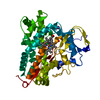
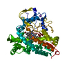
| Structure viewer | Molecule: MolmilJmol/JSmol |
|---|
- Downloads & links
-Download
| PDBx/mmCIF format | 4l7p.cif.gz | 150.4 KB | Display | PDBx/mmCIF format |
|---|---|---|---|---|
| PDB format | pdb4l7p.ent.gz | 117 KB | Display | PDB format |
| PDBx/mmJSON format | 4l7p.json.gz | Tree view | PDBx/mmJSON format | |
| Others |  Other downloads Other downloads |
-Validation report
| Arichive directory | https://data.pdbj.org/pub/pdb/validation_reports/l7/4l7pftp://data.pdbj.org/pub/pdb/validation_reports/l7/4l7p | HTTPS FTP |
|---|
-Related structure data
| Related structure data |  4l6zC  4l70C  4l7lC  4l7nC  4l7oC  4l7rC  4l7uC  4gv4S C: citing same article ( S: Starting model for refinement |
|---|---|
| Similar structure data |


-Links
 PDBj
PDBj- Assembly
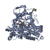
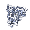
| Deposited unit | 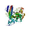
| ||||||||
|---|---|---|---|---|---|---|---|---|---|
| 1 |
| ||||||||
| Unit cell |
| ||||||||
| Details | MONOMERIC |
-Components
| #1: Protein | Mass: 39752.074 Da / Num. of mol.: 1 / Fragment: CATALYTIC PARP DOMAIN Source method: isolated from a genetically manipulated source Source: (gene. exp.) Homo sapiens (human) / Gene: ADPRT3, ADPRTL3, PARP3 / Plasmid: PNIC-BSA4 / Production host:  Escherichia coli (E. coli) / Strain (production host): BL21 (DE3) R3 PRARE / References: UniProt: Q9Y6F1, NAD+ ADP-ribosyltransferase Escherichia coli (E. coli) / Strain (production host): BL21 (DE3) R3 PRARE / References: UniProt: Q9Y6F1, NAD+ ADP-ribosyltransferase |
|---|---|
| #2: Chemical | ChemComp-M95 / (  Mass: 349.383 Da / Num. of mol.: 1 / Source method: obtained synthetically / Formula: C20H19N3O3 Mass: 349.383 Da / Num. of mol.: 1 / Source method: obtained synthetically / Formula: C20H19N3O3 |
| #3: Water | ChemComp-HOH / Water Mass: 18.015 Da / Num. of mol.: 46 / Source method: isolated from a natural source / Formula: H2O Mass: 18.015 Da / Num. of mol.: 46 / Source method: isolated from a natural source / Formula: H2O |
-Experimental details
-Experiment
| Experiment | Method: X-RAY DIFFRACTION / Number of used crystals: 1 |
|---|
- Sample preparation
| Crystal | Density Matthews: 2.16 Å3/Da / Density % sol: 43.1 % |
|---|---|
| Crystal grow | Temperature: 293 K / Method: vapor diffusion, sitting drop / pH: 7 Details: 1.9M DL-Malic Acid, 0.1M Bis-tris-propane , pH 7.0, VAPOR DIFFUSION, SITTING DROP, temperature 293K |
-Data collection
| Diffraction | Mean temperature: 100 K |
|---|---|
| Diffraction source | Source: SYNCHROTRON / Site: BESSY  / Beamline: 14.1 / Wavelength: 0.91841 Å / Beamline: 14.1 / Wavelength: 0.91841 Å |
| Detector | Type: RAYONIX MX-225 / Detector: CCD / Date: Feb 2, 2012 |
| Radiation | Monochromator: Si 111 / Protocol: SINGLE WAVELENGTH / Monochromatic (M) / Laue (L): M / Scattering type: x-ray |
| Radiation wavelength | Wavelength: 0.91841 Å / Relative weight: 1 |
| Reflection | Resolution: 2.3→30 Å / Num. all: 15221 / Num. obs: 15221 / % possible obs: 99.5 % / Observed criterion σ(F): 0 / Observed criterion σ(I): 0 / Redundancy: 5.7 % / Biso Wilson estimate: 40.27 Å2 / Rmerge(I) obs: 0.076 / Rsym value: 0.106 / Net I/σ(I): 19.6 |
| Reflection shell | Resolution: 2.3→2.36 Å / Redundancy: 5.7 % / Rmerge(I) obs: 0.603 / Mean I/σ(I) obs: 3.2 / Num. unique all: 1133 / Rsym value: 0.523 / % possible all: 99.7 |
- Processing
| Software |
| ||||||||||||||||||||||||||||||||||||||||||||||||||||||||||||||||||||||||||||||||||||||||||||||||||||||||||||||||||
|---|---|---|---|---|---|---|---|---|---|---|---|---|---|---|---|---|---|---|---|---|---|---|---|---|---|---|---|---|---|---|---|---|---|---|---|---|---|---|---|---|---|---|---|---|---|---|---|---|---|---|---|---|---|---|---|---|---|---|---|---|---|---|---|---|---|---|---|---|---|---|---|---|---|---|---|---|---|---|---|---|---|---|---|---|---|---|---|---|---|---|---|---|---|---|---|---|---|---|---|---|---|---|---|---|---|---|---|---|---|---|---|---|---|---|---|
| Refinement | Method to determine structure: MOLECULAR REPLACEMENT Starting model: PDB ENTRY 4GV4 Resolution: 2.3→20.76 Å / Cor.coef. Fo:Fc: 0.9301 / Cor.coef. Fo:Fc free: 0.907 / SU R Cruickshank DPI: 0.35 / Cross valid method: THROUGHOUT / σ(F): 0
| ||||||||||||||||||||||||||||||||||||||||||||||||||||||||||||||||||||||||||||||||||||||||||||||||||||||||||||||||||
| Displacement parameters | Biso mean: 42.57 Å2
| ||||||||||||||||||||||||||||||||||||||||||||||||||||||||||||||||||||||||||||||||||||||||||||||||||||||||||||||||||
| Refine analyze | Luzzati coordinate error obs: 0.251 Å | ||||||||||||||||||||||||||||||||||||||||||||||||||||||||||||||||||||||||||||||||||||||||||||||||||||||||||||||||||
| Refinement step | Cycle: LAST / Resolution: 2.3→20.76 Å
| ||||||||||||||||||||||||||||||||||||||||||||||||||||||||||||||||||||||||||||||||||||||||||||||||||||||||||||||||||
| Refine LS restraints |
| ||||||||||||||||||||||||||||||||||||||||||||||||||||||||||||||||||||||||||||||||||||||||||||||||||||||||||||||||||
| LS refinement shell | Resolution: 2.3→2.46 Å / Total num. of bins used: 8
| ||||||||||||||||||||||||||||||||||||||||||||||||||||||||||||||||||||||||||||||||||||||||||||||||||||||||||||||||||
| Refinement TLS params. | Method: refined / Origin x: 2.222 Å / Origin y: -0.5828 Å / Origin z: 22.849 Å
| ||||||||||||||||||||||||||||||||||||||||||||||||||||||||||||||||||||||||||||||||||||||||||||||||||||||||||||||||||
| Refinement TLS group | Selection details: { A|177 - A|532 } |
