 Movie
Movie Controller

[English] 日本語
 Yorodumi
- PDB-4l6e: Crystal Structure of the RanBD1 fourth domain of E3 SUMO-protein ... -
+ Open data

- Basic information
| Entry | Database: PDB / ID: 4l6e | ||||||
|---|---|---|---|---|---|---|---|
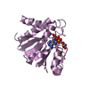
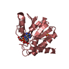
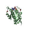
| Title | Crystal Structure of the RanBD1 fourth domain of E3 SUMO-protein ligase RanBP2. Northeast Structural Genomics Consortium (NESG) Target HR9193b | ||||||
 Components Components | E3 SUMO-protein ligase RanBP2 | ||||||
 Keywords Keywords |  LIGASE / ISOMERASE / Structural Genomics / PSI-Biology / Protein Structure Initiative / NESG / RanBP2 / RanBD1 / Northeast Structural Genomics Consortium LIGASE / ISOMERASE / Structural Genomics / PSI-Biology / Protein Structure Initiative / NESG / RanBP2 / RanBD1 / Northeast Structural Genomics Consortium | ||||||
| Function / homology |  Function and homology information Function and homology informationcytoplasmic periphery of the nuclear pore complex / SUMO ligase activity / SUMO ligase complex / annulate lamellae / nuclear pore cytoplasmic filaments / Nuclear Pore Complex (NPC) Disassembly / nuclear inclusion body / nuclear pore nuclear basket / Transport of Ribonucleoproteins into the Host Nucleus / Regulation of Glucokinase by Glucokinase Regulatory Protein ...cytoplasmic periphery of the nuclear pore complex / SUMO ligase activity / SUMO ligase complex / annulate lamellae / nuclear pore cytoplasmic filaments / Nuclear Pore Complex (NPC) Disassembly / nuclear inclusion body / nuclear pore nuclear basket / Transport of Ribonucleoproteins into the Host Nucleus / Regulation of Glucokinase by Glucokinase Regulatory Protein / Defective TPR may confer susceptibility towards thyroid papillary carcinoma (TPC) / Transport of the SLBP independent Mature mRNA / Transport of the SLBP Dependant Mature mRNA / NS1 Mediated Effects on Host Pathways / SUMOylation of SUMOylation proteins / Transport of Mature mRNA Derived from an Intronless Transcript / Transferases; Acyltransferases; Aminoacyltransferases / Rev-mediated nuclear export of HIV RNA / SUMOylation of RNA binding proteins / nuclear export / Nuclear import of Rev protein / Transport of Mature mRNA derived from an Intron-Containing Transcript / NEP/NS2 Interacts with the Cellular Export Machinery / tRNA processing in the nucleus / SUMO transferase activity / nucleocytoplasmic transport / centrosome localization / Viral Messenger RNA Synthesis / regulation of gluconeogenesis / NLS-bearing protein import into nucleus / SUMOylation of ubiquitinylation proteins / Vpr-mediated nuclear import of PICs / SUMOylation of DNA replication proteins / protein sumoylation / Regulation of HSF1-mediated heat shock response / mRNA transport / Amplification of signal from unattached kinetochores via a MAD2 inhibitory signal / SUMOylation of DNA damage response and repair proteins / nuclear pore / Mitotic Prometaphase / EML4 and NUDC in mitotic spindle formation / Resolution of Sister Chromatid Cohesion / response to amphetamine / SUMOylation of chromatin organization proteins / GTPase activator activity / HCMV Late Events / RHO GTPases Activate Formins / Transcriptional regulation by small RNAs / Signaling by ALK fusions and activated point mutants / ISG15 antiviral mechanism / small GTPase binding / HCMV Early Events / Separation of Sister Chromatids / protein folding / nuclear envelope / snRNP Assembly / nuclear membrane / intracellular membrane-bounded organelle / protein-containing complex binding / SARS-CoV-2 activates/modulates innate and adaptive immune responses / RNA binding / nucleoplasm / membrane / metal ion binding / cytosol / cytoplasmSimilarity search - Function | ||||||
| Biological species |  Homo sapiens (human) Homo sapiens (human) | ||||||
| Method | X-RAY DIFFRACTION / SYNCHROTRON / SAD / Resolution: 2.496 Å | ||||||
 Authors Authors | Vorobiev, S. / Su, M. / Seetharaman, J. / Mao, L. / Xiao, R. / Maglaqui, M. / Kogan, S. / Wang, H. / Everett, J.K. / Acton, T.B. ...Vorobiev, S. / Su, M. / Seetharaman, J. / Mao, L. / Xiao, R. / Maglaqui, M. / Kogan, S. / Wang, H. / Everett, J.K. / Acton, T.B. / Montelione, G.T. / Hunt, J.F. / Tong, L. / Northeast Structural Genomics Consortium (NESG) | ||||||
 Citation Citation | Journal: To be Published Title: Crystal Structure of the RanBD1 fourth domain of E3 SUMO-protein ligase RanBP2. Authors: Vorobiev, S. / Su, M. / Seetharaman, J. / Mao, L. / Xiao, R. / Maglaqui, M. / Kogan, S. / Wang, H. / Everett, J.K. / Acton, T.B. / Montelione, G.T. / Hunt, J.F. / Tong, L. | ||||||
| History |
|
- Structure visualization
| Structure viewer | Molecule: MolmilJmol/JSmol |
|---|
- Downloads & links
-Download
| PDBx/mmCIF format | 4l6e.cif.gz | 63.7 KB | Display | PDBx/mmCIF format |
|---|---|---|---|---|
| PDB format | pdb4l6e.ent.gz | 51 KB | Display | PDB format |
| PDBx/mmJSON format | 4l6e.json.gz | Tree view | PDBx/mmJSON format | |
| Others |  Other downloads Other downloads |
-Validation report
| Arichive directory | https://data.pdbj.org/pub/pdb/validation_reports/l6/4l6eftp://data.pdbj.org/pub/pdb/validation_reports/l6/4l6e | HTTPS FTP |
|---|
-Related structure data
| Similar structure data | |
|---|---|
| Other databases |


-Links
 PDBj
PDBj


- Assembly
| Deposited unit | 
| |||||||||
|---|---|---|---|---|---|---|---|---|---|---|
| 1 |
| |||||||||
| Unit cell |
| |||||||||
| Components on special symmetry positions |
|
-Components
| #1: Protein | Mass: 18708.486 Da / Num. of mol.: 1 / Fragment: RanBD1 4 domain (UNP residues 2907-3050) Source method: isolated from a genetically manipulated source Source: (gene. exp.) Homo sapiens (human) / Gene: NUP358, RANBP2 / Plasmid: HR9193B-2907-3050-15.1 / Production host:  Escherichia coli (E. coli) / Strain (production host): BL21(DE3)+ Magic Escherichia coli (E. coli) / Strain (production host): BL21(DE3)+ MagicReferences: UniProt: P49792, Ligases; Forming carbon-nitrogen bonds; Acid-amino-acid ligases (peptide synthases), peptidylprolyl isomerase |
|---|---|
| #2: Water | ChemComp-HOH / Water Mass: 18.015 Da / Num. of mol.: 37 / Source method: isolated from a natural source / Formula: H2O Mass: 18.015 Da / Num. of mol.: 37 / Source method: isolated from a natural source / Formula: H2O |
-Experimental details
-Experiment
| Experiment | Method: X-RAY DIFFRACTION / Number of used crystals: 1 |
|---|
- Sample preparation
| Crystal | Density Matthews: 2.84 Å3/Da / Density % sol: 56.7 % |
|---|---|
| Crystal grow | Temperature: 277 K / Method: microbatch crystallization under oil / pH: 7 Details: 12% PEG 3350, 0.1M sodium acetate, pH 7.00, Microbatch crystallization under oil, temperature 277K |
-Data collection
| Diffraction | Mean temperature: 100 K |
|---|---|
| Diffraction source | Source: SYNCHROTRON / Site: NSLS  / Beamline: X4C / Wavelength: 0.97924 Å / Beamline: X4C / Wavelength: 0.97924 Å |
| Detector | Type: MAR CCD 165 mm / Detector: CCD / Date: Jun 5, 2013 |
| Radiation | Monochromator: Si 111 CHANNEL / Protocol: SINGLE WAVELENGTH / Monochromatic (M) / Laue (L): M / Scattering type: x-ray |
| Radiation wavelength | Wavelength: 0.97924 Å / Relative weight: 1 |
| Reflection | Resolution: 2.5→50 Å / Num. obs: 14342 / % possible obs: 99.5 % / Observed criterion σ(F): 0 / Observed criterion σ(I): 0 / Redundancy: 2 % / Biso Wilson estimate: 69.21 Å2 / Rmerge(I) obs: 0.064 / Net I/σ(I): 16 |
| Reflection shell | Resolution: 2.5→2.59 Å / Redundancy: 2 % / Rmerge(I) obs: 0.558 / Mean I/σ(I) obs: 0.98 / Num. unique all: 1645 / % possible all: 100 |
- Processing
| Software |
| ||||||||||||||||||||||||||||||||||||||||||
|---|---|---|---|---|---|---|---|---|---|---|---|---|---|---|---|---|---|---|---|---|---|---|---|---|---|---|---|---|---|---|---|---|---|---|---|---|---|---|---|---|---|---|---|
| Refinement | Method to determine structure: SAD / Resolution: 2.496→32.212 Å / Occupancy max: 1 / Occupancy min: 0.5 / SU ML: 0.79 / Isotropic thermal model: anisotropic / Cross valid method: THROUGHOUT / σ(F): 1.09 / Phase error: 27.34 / Stereochemistry target values: ML
| ||||||||||||||||||||||||||||||||||||||||||
| Solvent computation | Shrinkage radii: 0.86 Å / VDW probe radii: 1.1 Å / Solvent model: FLAT BULK SOLVENT MODEL / Bsol: 64.205 Å2 / ksol: 0.338 e/Å3 | ||||||||||||||||||||||||||||||||||||||||||
| Displacement parameters | Biso max: 183.91 Å2 / Biso mean: 79.441 Å2 / Biso min: 46.25 Å2
| ||||||||||||||||||||||||||||||||||||||||||
| Refinement step | Cycle: LAST / Resolution: 2.496→32.212 Å
| ||||||||||||||||||||||||||||||||||||||||||
| Refine LS restraints |
| ||||||||||||||||||||||||||||||||||||||||||
| LS refinement shell | Refine-ID: X-RAY DIFFRACTION / Total num. of bins used: 5
| ||||||||||||||||||||||||||||||||||||||||||
| Refinement TLS params. | Method: refined / Origin x: 15.9328 Å / Origin y: 19.9245 Å / Origin z: 74.7614 Å
| ||||||||||||||||||||||||||||||||||||||||||
| Refinement TLS group | Selection details: chain A |
