 Movie
Movie Controller

[English] 日本語
 Yorodumi
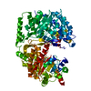
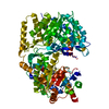
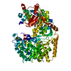
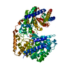
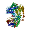
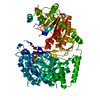
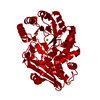
- PDB-4l65: Crystal structure of the Candida albicans Methionine Synthase in ... -
+ Open data

- Basic information
| Entry | Database: PDB / ID: 4l65 | ||||||
|---|---|---|---|---|---|---|---|
| Title | Crystal structure of the Candida albicans Methionine Synthase in complex with 5-Methyl-Tetrahydrofolate and Methionine | ||||||
 Components Components | 5-methyltetrahydropteroyltriglutamate--homocysteine methyltransferase | ||||||
 Keywords Keywords |  TRANSFERASE / cobalamin-independent / surface entropy reduction / fungal / Dual TIM Barrels / Methionine synthase TRANSFERASE / cobalamin-independent / surface entropy reduction / fungal / Dual TIM Barrels / Methionine synthase | ||||||
| Function / homology |  Function and homology information Function and homology informationL-methionine biosynthetic process from homoserine via O-acetyl-L-homoserine and cystathionine / : / 5-methyltetrahydropteroyltriglutamate-homocysteine S-methyltransferase / 5-methyltetrahydropteroyltriglutamate-homocysteine S-methyltransferase activity / fungal biofilm matrix / hyphal cell wall / adenine biosynthetic process / methionine metabolic process / 'de novo' L-methionine biosynthetic process / fungal-type cell wall ...L-methionine biosynthetic process from homoserine via O-acetyl-L-homoserine and cystathionine / : / 5-methyltetrahydropteroyltriglutamate-homocysteine S-methyltransferase / 5-methyltetrahydropteroyltriglutamate-homocysteine S-methyltransferase activity / fungal biofilm matrix / hyphal cell wall / adenine biosynthetic process / methionine metabolic process / 'de novo' L-methionine biosynthetic process / fungal-type cell wall / methionine biosynthetic process / cellular response to heat / methylation / cell surface / zinc ion binding / nucleusSimilarity search - Function | ||||||
| Biological species |  Candida albicans SC5314 (yeast) Candida albicans SC5314 (yeast) | ||||||
| Method | X-RAY DIFFRACTION / SYNCHROTRON / MOLECULAR REPLACEMENT / Resolution: 2.31 Å | ||||||
 Authors Authors | Ubhi, D. / Robertus, J.D. | ||||||
 Citation Citation | Journal: J.Mol.Biol. / Year: 2014 Title: Structural analysis of a fungal methionine synthase with substrates and inhibitors. Authors: Ubhi, D. / Kago, G. / Monzingo, A.F. / Robertus, J.D. | ||||||
| History |
|
- Structure visualization
| Structure viewer | Molecule: MolmilJmol/JSmol |
|---|
- Downloads & links
-Download
| PDBx/mmCIF format | 4l65.cif.gz | 301.2 KB | Display | PDBx/mmCIF format |
|---|---|---|---|---|
| PDB format | pdb4l65.ent.gz | 241.5 KB | Display | PDB format |
| PDBx/mmJSON format | 4l65.json.gz | Tree view | PDBx/mmJSON format | |
| Others |  Other downloads Other downloads |
-Validation report
| Arichive directory | https://data.pdbj.org/pub/pdb/validation_reports/l6/4l65ftp://data.pdbj.org/pub/pdb/validation_reports/l6/4l65 | HTTPS FTP |
|---|
-Related structure data


-Links
 PDBj
PDBj- Assembly
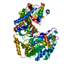
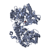
| Deposited unit | 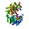
| ||||||||
|---|---|---|---|---|---|---|---|---|---|
| 1 |
| ||||||||
| Unit cell |
|
-Components
| #1: Protein | Mass: 88188.961 Da / Num. of mol.: 1 / Mutation: K103A, K104A, E107A Source method: isolated from a genetically manipulated source Source: (gene. exp.) Candida albicans SC5314 (yeast) / Strain: BWP17 / Gene: CaO19.10083, CaO19.2551, MET6 / Plasmid: pNIC28-Bsa4 / Production host:  Escherichia coli (E. coli) / Strain (production host): BL21(DE3) Escherichia coli (E. coli) / Strain (production host): BL21(DE3)References: UniProt: P82610, 5-methyltetrahydropteroyltriglutamate-homocysteine S-methyltransferase |
|---|---|
| #2: Chemical | ChemComp-ZN /   Mass: 65.409 Da / Num. of mol.: 1 / Source method: obtained synthetically / Formula: Zn Mass: 65.409 Da / Num. of mol.: 1 / Source method: obtained synthetically / Formula: Zn |
| #3: Chemical | ChemComp-C2F / Levomefolic acid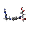  Mass: 459.456 Da / Num. of mol.: 1 / Source method: obtained synthetically / Formula: C20H25N7O6 Mass: 459.456 Da / Num. of mol.: 1 / Source method: obtained synthetically / Formula: C20H25N7O6 |
| #4: Chemical | ChemComp-MET / Methionine  Type: L-peptide linking / Mass: 149.211 Da / Num. of mol.: 1 / Source method: obtained synthetically / Formula: C5H11NO2S Type: L-peptide linking / Mass: 149.211 Da / Num. of mol.: 1 / Source method: obtained synthetically / Formula: C5H11NO2S |
| #5: Water | ChemComp-HOH / Water Mass: 18.015 Da / Num. of mol.: 137 / Source method: isolated from a natural source / Formula: H2O Mass: 18.015 Da / Num. of mol.: 137 / Source method: isolated from a natural source / Formula: H2O |
-Experimental details
-Experiment
| Experiment | Method: X-RAY DIFFRACTION / Number of used crystals: 1 |
|---|
- Sample preparation
| Crystal | Density Matthews: 2.18 Å3/Da / Density % sol: 43.49 % |
|---|---|
| Crystal grow | Temperature: 293 K / Method: vapor diffusion, hanging drop / pH: 7.4 Details: 50 mM NaI, 27% (w/v) PEG 3350, 0.25 mM DTT, 0.15 mM ZnSO4, 5 mM 5-Methyl-Tetrahydrofolate-Glu3, 10 mM Methionine, 20 mM Tris-Cl pH 7.4 , VAPOR DIFFUSION, HANGING DROP, temperature 293K |
-Data collection
| Diffraction | Mean temperature: 100 K |
|---|---|
| Diffraction source | Source: SYNCHROTRON / Site: ALS  / Beamline: 5.0.3 / Wavelength: 0.97648 Å / Beamline: 5.0.3 / Wavelength: 0.97648 Å |
| Detector | Type: ADSC QUANTUM 315r / Detector: CCD / Date: May 13, 2012 / Details: Asymmetric cut single crystal Si (220) |
| Radiation | Monochromator: Asymmetric cut single crystal Si (220) / Protocol: SINGLE WAVELENGTH / Monochromatic (M) / Laue (L): M / Scattering type: x-ray |
| Radiation wavelength | Wavelength: 0.97648 Å / Relative weight: 1 |
| Reflection | Resolution: 2.3→49.52 Å / Num. all: 34148 / Num. obs: 34134 / % possible obs: 100 % / Observed criterion σ(I): 0 / Redundancy: 7.2 % / Rmerge(I) obs: 0.066 / Net I/σ(I): 11 |
| Reflection shell | Resolution: 2.33→2.37 Å / Rmerge(I) obs: 0.549 / % possible all: 99.8 |
- Processing
| Software |
| |||||||||||||||||||||||||||||||||||||||||||||||||||||||||||||||||||||||||||||||||||||||||||||||||||||||||||||||||||||||||||||||||||||||||||||||||||||||||||||||||||||||||||||||
|---|---|---|---|---|---|---|---|---|---|---|---|---|---|---|---|---|---|---|---|---|---|---|---|---|---|---|---|---|---|---|---|---|---|---|---|---|---|---|---|---|---|---|---|---|---|---|---|---|---|---|---|---|---|---|---|---|---|---|---|---|---|---|---|---|---|---|---|---|---|---|---|---|---|---|---|---|---|---|---|---|---|---|---|---|---|---|---|---|---|---|---|---|---|---|---|---|---|---|---|---|---|---|---|---|---|---|---|---|---|---|---|---|---|---|---|---|---|---|---|---|---|---|---|---|---|---|---|---|---|---|---|---|---|---|---|---|---|---|---|---|---|---|---|---|---|---|---|---|---|---|---|---|---|---|---|---|---|---|---|---|---|---|---|---|---|---|---|---|---|---|---|---|---|---|---|---|
| Refinement | Method to determine structure: MOLECULAR REPLACEMENT / Resolution: 2.31→49.52 Å / Cor.coef. Fo:Fc: 0.949 / Cor.coef. Fo:Fc free: 0.903 / SU B: 16.051 / SU ML: 0.197 / Cross valid method: THROUGHOUT / ESU R: 0.381 / ESU R Free: 0.274 / Stereochemistry target values: MAXIMUM LIKELIHOOD / Details: HYDROGENS HAVE BEEN ADDED IN THE RIDING POSITIONS
| |||||||||||||||||||||||||||||||||||||||||||||||||||||||||||||||||||||||||||||||||||||||||||||||||||||||||||||||||||||||||||||||||||||||||||||||||||||||||||||||||||||||||||||||
| Solvent computation | Ion probe radii: 0.8 Å / Shrinkage radii: 0.8 Å / VDW probe radii: 1.2 Å / Solvent model: MASK | |||||||||||||||||||||||||||||||||||||||||||||||||||||||||||||||||||||||||||||||||||||||||||||||||||||||||||||||||||||||||||||||||||||||||||||||||||||||||||||||||||||||||||||||
| Displacement parameters | Biso mean: 56.832 Å2
| |||||||||||||||||||||||||||||||||||||||||||||||||||||||||||||||||||||||||||||||||||||||||||||||||||||||||||||||||||||||||||||||||||||||||||||||||||||||||||||||||||||||||||||||
| Refinement step | Cycle: LAST / Resolution: 2.31→49.52 Å
| |||||||||||||||||||||||||||||||||||||||||||||||||||||||||||||||||||||||||||||||||||||||||||||||||||||||||||||||||||||||||||||||||||||||||||||||||||||||||||||||||||||||||||||||
| Refine LS restraints |
| |||||||||||||||||||||||||||||||||||||||||||||||||||||||||||||||||||||||||||||||||||||||||||||||||||||||||||||||||||||||||||||||||||||||||||||||||||||||||||||||||||||||||||||||
| LS refinement shell | Resolution: 2.314→2.374 Å / Total num. of bins used: 20
| |||||||||||||||||||||||||||||||||||||||||||||||||||||||||||||||||||||||||||||||||||||||||||||||||||||||||||||||||||||||||||||||||||||||||||||||||||||||||||||||||||||||||||||||
| Refinement TLS params. | Method: refined / Refine-ID: X-RAY DIFFRACTION
| |||||||||||||||||||||||||||||||||||||||||||||||||||||||||||||||||||||||||||||||||||||||||||||||||||||||||||||||||||||||||||||||||||||||||||||||||||||||||||||||||||||||||||||||
| Refinement TLS group |
|
