 Movie
Movie Controller


+ Open data

- Basic information
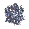
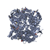
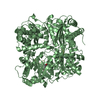
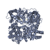
| Entry | Database: PDB / ID: 4l3t | ||||||
|---|---|---|---|---|---|---|---|
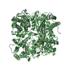
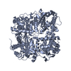
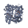
| Title | Crystal Structure of Substrate-free Human Presequence Protease | ||||||
 Components Components | (Presequence protease, ...) x 2 | ||||||
 Keywords Keywords |  HYDROLASE / zinc metalloendoprotease / mitochondrial matrix HYDROLASE / zinc metalloendoprotease / mitochondrial matrix | ||||||
| Function / homology |  Function and homology informationMitochondrial protein import / protein targeting to mitochondrion / Hydrolases; Acting on peptide bonds (peptidases); Metalloendopeptidases / enzyme activator activity / protein processing / metalloendopeptidase activity / metallopeptidase activity / mitochondrial matrix / mitochondrion / proteolysis / zinc ion binding Function and homology informationMitochondrial protein import / protein targeting to mitochondrion / Hydrolases; Acting on peptide bonds (peptidases); Metalloendopeptidases / enzyme activator activity / protein processing / metalloendopeptidase activity / metallopeptidase activity / mitochondrial matrix / mitochondrion / proteolysis / zinc ion bindingSimilarity search - Function | ||||||
| Biological species |  Homo sapiens (human) Homo sapiens (human) | ||||||
| Method | X-RAY DIFFRACTION / SYNCHROTRON / molecular replacement/SAD / Resolution: 2.03 Å | ||||||
 Authors Authors | King, J.V. / Liang, W.G. / Tang, W.J. | ||||||
 Citation Citation | Journal: Structure / Year: 2014 Title: Molecular basis of substrate recognition and degradation by human presequence protease. Authors: King, J.V. / Liang, W.G. / Scherpelz, K.P. / Schilling, A.B. / Meredith, S.C. / Tang, W.J. | ||||||
| History |
|
- Structure visualization
| Structure viewer | Molecule: MolmilJmol/JSmol |
|---|
- Downloads & links
-Download
| PDBx/mmCIF format | 4l3t.cif.gz | 830.9 KB | Display | PDBx/mmCIF format |
|---|---|---|---|---|
| PDB format | pdb4l3t.ent.gz | 702.9 KB | Display | PDB format |
| PDBx/mmJSON format | 4l3t.json.gz | Tree view | PDBx/mmJSON format | |
| Others |  Other downloads Other downloads |
-Validation report
| Arichive directory | https://data.pdbj.org/pub/pdb/validation_reports/l3/4l3tftp://data.pdbj.org/pub/pdb/validation_reports/l3/4l3t | HTTPS FTP |
|---|
-Related structure data


-Links
 PDBj
PDBj

- Assembly
| Deposited unit | 
| ||||||||
|---|---|---|---|---|---|---|---|---|---|
| 1 | 
| ||||||||
| 2 | 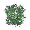
| ||||||||
| Unit cell |
|
-Components
-Presequence protease, ... , 2 types, 2 molecules AB
| #1: Protein | Mass: 116454.930 Da / Num. of mol.: 1 / Fragment: UNP residues 33-1037 / Mutation: E107Q Source method: isolated from a genetically manipulated source Source: (gene. exp.) Homo sapiens (human) / Gene: KIAA1104, MP1, PITRM1 / Plasmid: pProExH6 / Production host:  Escherichia coli (E. coli) / Strain (production host): Rosetta(DE3) Escherichia coli (E. coli) / Strain (production host): Rosetta(DE3)References: UniProt: Q5JRX3, Hydrolases; Acting on peptide bonds (peptidases); Metalloendopeptidases |
|---|---|
| #2: Protein | Mass: 116465.914 Da / Num. of mol.: 1 / Fragment: UNP residues 33-1037 / Mutation: E107Q Source method: isolated from a genetically manipulated source Source: (gene. exp.) Homo sapiens (human) / Gene: KIAA1104, MP1, PITRM1 / Plasmid: pProExH6 / Production host: Escherichia coli (E. coli) / Strain (production host): Rosetta(DE3)References: UniProt: Q5JRX3, Hydrolases; Acting on peptide bonds (peptidases); Metalloendopeptidases |
-Non-polymers , 4 types, 1080 molecules 


| #3: Chemical |  Mass: 65.409 Da / Num. of mol.: 2 / Source method: obtained synthetically / Formula: Zn Mass: 65.409 Da / Num. of mol.: 2 / Source method: obtained synthetically / Formula: Zn#4: Chemical | ChemComp-GOL / Glycerol Mass: 92.094 Da / Num. of mol.: 7 / Source method: obtained synthetically / Formula: C3H8O3 Mass: 92.094 Da / Num. of mol.: 7 / Source method: obtained synthetically / Formula: C3H8O3#5: Chemical | ChemComp-ACT / Acetate Mass: 59.044 Da / Num. of mol.: 13 / Source method: obtained synthetically / Formula: C2H3O2 Mass: 59.044 Da / Num. of mol.: 13 / Source method: obtained synthetically / Formula: C2H3O2#6: Water | ChemComp-HOH / | WaterMass: 18.015 Da / Num. of mol.: 1058 / Source method: isolated from a natural source / Formula: H2O |
|---|
-Details
| Sequence details | I328V, A397V, AND Q1037R ARE NATURAL VARIANTS. |
|---|
-Experimental details
-Experiment
| Experiment | Method: X-RAY DIFFRACTION / Number of used crystals: 1 |
|---|
- Sample preparation
| Crystal | Density Matthews: 2.82 Å3/Da / Density % sol: 56.39 % |
|---|---|
| Crystal grow | Temperature: 291.15 K / Method: vapor diffusion, hanging drop / pH: 6.5 Details: 15.2% w/v PEG8000, 15 mM TCEP, 80 mM sodium cacodylate, pH 6.5, 160 mM calcium acetate, 20% v/v glycerol, VAPOR DIFFUSION, HANGING DROP, temperature 291.15K |
-Data collection
| Diffraction | Mean temperature: 113.5 K |
|---|---|
| Diffraction source | Source: SYNCHROTRON / Site: APS  / Beamline: 19-ID / Wavelength: 0.97895 Å / Beamline: 19-ID / Wavelength: 0.97895 Å |
| Detector | Type: ADSC QUANTUM 315r / Detector: CCD / Date: Oct 15, 2012 |
| Radiation | Monochromator: Rosenbaum-Rock high-resolution double-crystal Si(111) Protocol: SINGLE WAVELENGTH / Monochromatic (M) / Laue (L): M / Scattering type: x-ray |
| Radiation wavelength | Wavelength: 0.97895 Å / Relative weight: 1 |
| Reflection | Resolution: 2.03→50 Å / Num. all: 166830 / Num. obs: 163827 / % possible obs: 98.2 % / Observed criterion σ(F): 0 / Observed criterion σ(I): -3 / Redundancy: 4.13 % / Biso Wilson estimate: 27.23 Å2 / Rsym value: 0.086 / Net I/σ(I): 17.1 |
| Reflection shell | Resolution: 2.03→2.07 Å / Redundancy: 3.8 % / Mean I/σ(I) obs: 2.75 / Num. unique all: 8053 / Rsym value: 0.585 / % possible all: 97 |
- Processing
| Software |
| |||||||||||||||||||||||||||||||||||||||||||||||||||||||||||||||||||||||||||||||||||||||||||||||||||||||||||||||||||||||||||||||||||||||||||||||||||||||||||||||||||||||||||||||||||||||||||||||||||||||||||||||||||||||||
|---|---|---|---|---|---|---|---|---|---|---|---|---|---|---|---|---|---|---|---|---|---|---|---|---|---|---|---|---|---|---|---|---|---|---|---|---|---|---|---|---|---|---|---|---|---|---|---|---|---|---|---|---|---|---|---|---|---|---|---|---|---|---|---|---|---|---|---|---|---|---|---|---|---|---|---|---|---|---|---|---|---|---|---|---|---|---|---|---|---|---|---|---|---|---|---|---|---|---|---|---|---|---|---|---|---|---|---|---|---|---|---|---|---|---|---|---|---|---|---|---|---|---|---|---|---|---|---|---|---|---|---|---|---|---|---|---|---|---|---|---|---|---|---|---|---|---|---|---|---|---|---|---|---|---|---|---|---|---|---|---|---|---|---|---|---|---|---|---|---|---|---|---|---|---|---|---|---|---|---|---|---|---|---|---|---|---|---|---|---|---|---|---|---|---|---|---|---|---|---|---|---|---|---|---|---|---|---|---|---|---|---|---|---|---|---|---|---|---|
| Refinement | Method to determine structure: molecular replacement/SAD / Resolution: 2.03→42.547 Å / SU ML: 0.19 / σ(F): 0 / Phase error: 21.36 / Stereochemistry target values: ML Details: THE PRESENCE IN THE ASYMMETRIC UNIT OF TWO PROTEINS WITH DISTINCT PATTERNS OF SIDE CHAIN MODIFICATION REPRESENTS THE BEST FIT TO THE ELECTRON DENSITY AND IS NOT DERIVED FROM THE CO- ...Details: THE PRESENCE IN THE ASYMMETRIC UNIT OF TWO PROTEINS WITH DISTINCT PATTERNS OF SIDE CHAIN MODIFICATION REPRESENTS THE BEST FIT TO THE ELECTRON DENSITY AND IS NOT DERIVED FROM THE CO-CRYSTALLIZATION OF TWO CHEMICALLY DISTINCT PROTEINS.
| |||||||||||||||||||||||||||||||||||||||||||||||||||||||||||||||||||||||||||||||||||||||||||||||||||||||||||||||||||||||||||||||||||||||||||||||||||||||||||||||||||||||||||||||||||||||||||||||||||||||||||||||||||||||||
| Solvent computation | Shrinkage radii: 0.6 Å / VDW probe radii: 0.9 Å / Solvent model: FLAT BULK SOLVENT MODEL / Bsol: 53.486 Å2 / ksol: 0.38 e/Å3 | |||||||||||||||||||||||||||||||||||||||||||||||||||||||||||||||||||||||||||||||||||||||||||||||||||||||||||||||||||||||||||||||||||||||||||||||||||||||||||||||||||||||||||||||||||||||||||||||||||||||||||||||||||||||||
| Displacement parameters | Biso mean: 36.11 Å2
| |||||||||||||||||||||||||||||||||||||||||||||||||||||||||||||||||||||||||||||||||||||||||||||||||||||||||||||||||||||||||||||||||||||||||||||||||||||||||||||||||||||||||||||||||||||||||||||||||||||||||||||||||||||||||
| Refinement step | Cycle: LAST / Resolution: 2.03→42.547 Å
| |||||||||||||||||||||||||||||||||||||||||||||||||||||||||||||||||||||||||||||||||||||||||||||||||||||||||||||||||||||||||||||||||||||||||||||||||||||||||||||||||||||||||||||||||||||||||||||||||||||||||||||||||||||||||
| Refine LS restraints |
| |||||||||||||||||||||||||||||||||||||||||||||||||||||||||||||||||||||||||||||||||||||||||||||||||||||||||||||||||||||||||||||||||||||||||||||||||||||||||||||||||||||||||||||||||||||||||||||||||||||||||||||||||||||||||
| LS refinement shell |
| |||||||||||||||||||||||||||||||||||||||||||||||||||||||||||||||||||||||||||||||||||||||||||||||||||||||||||||||||||||||||||||||||||||||||||||||||||||||||||||||||||||||||||||||||||||||||||||||||||||||||||||||||||||||||
| Refinement TLS params. | Method: refined / Origin x: 84.7848 Å / Origin y: 56.4114 Å / Origin z: -38.4891 Å
| |||||||||||||||||||||||||||||||||||||||||||||||||||||||||||||||||||||||||||||||||||||||||||||||||||||||||||||||||||||||||||||||||||||||||||||||||||||||||||||||||||||||||||||||||||||||||||||||||||||||||||||||||||||||||
| Refinement TLS group | Selection details: all |
