 Movie
Movie Controller

[English] 日本語
 Yorodumi
- PDB-4kzf: The mechanism of the amidases: The effect of the mutation E142L i... -
+ Open data

- Basic information
| Entry | Database: PDB / ID: 4kzf | ||||||
|---|---|---|---|---|---|---|---|
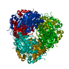
| Title | The mechanism of the amidases: The effect of the mutation E142L in the amidase from Geobacillus pallidus | ||||||
 Components Components | Aliphatic amidase | ||||||
 Keywords Keywords |  HYDROLASE / active site / chloride ion / cysteine 166 oxidation / amidase HYDROLASE / active site / chloride ion / cysteine 166 oxidation / amidase | ||||||
| Function / homology |  Function and homology information Function and homology information | ||||||
| Biological species |  | ||||||
| Method | X-RAY DIFFRACTION / SYNCHROTRON / MOLECULAR REPLACEMENT / molecular replacement / Resolution: 1.85 Å | ||||||
 Authors Authors | Weber, B.W. / Sewell, B.T. / Kimani, S.W. / Varsani, A. / Cowan, D.A. / Hunter, R. | ||||||
 Citation Citation | Journal: J.Biol.Chem. / Year: 2013 Title: The mechanism of the amidases: mutating the glutamate adjacent to the catalytic triad inactivates the enzyme due to substrate mispositioning. Authors: Weber, B.W. / Kimani, S.W. / Varsani, A. / Cowan, D.A. / Hunter, R. / Venter, G.A. / Gumbart, J.C. / Sewell, B.T. | ||||||
| History |
|
- Structure visualization
| Structure viewer | Molecule: MolmilJmol/JSmol |
|---|
- Downloads & links
-Download
| PDBx/mmCIF format | 4kzf.cif.gz | 85 KB | Display | PDBx/mmCIF format |
|---|---|---|---|---|
| PDB format | pdb4kzf.ent.gz | 63.4 KB | Display | PDB format |
| PDBx/mmJSON format | 4kzf.json.gz | Tree view | PDBx/mmJSON format | |
| Others |  Other downloads Other downloads |
-Validation report
| Arichive directory | https://data.pdbj.org/pub/pdb/validation_reports/kz/4kzfftp://data.pdbj.org/pub/pdb/validation_reports/kz/4kzf | HTTPS FTP |
|---|
-Related structure data
| Related structure data |  4gylC 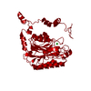 4gynC 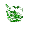 4lf0C  2plqS C: citing same article ( S: Starting model for refinement |
|---|---|
| Similar structure data |


-Links
 PDBj
PDBj- Assembly
| Deposited unit | 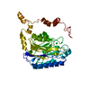
| ||||||||||||||||||||||||||||||
|---|---|---|---|---|---|---|---|---|---|---|---|---|---|---|---|---|---|---|---|---|---|---|---|---|---|---|---|---|---|---|---|
| 1 | x 6
| ||||||||||||||||||||||||||||||
| Unit cell |
| ||||||||||||||||||||||||||||||
| Components on special symmetry positions |
|
-Components
| #1: Protein | Mass: 38624.875 Da / Num. of mol.: 1 / Mutation: E142L Source method: isolated from a genetically manipulated source Source: (gene. exp.) Escherichia coli (E. coli) / Strain (production host): BL21(DE3) PLysS / References: UniProt: Q9L543, amidase |
|---|---|
| #2: Chemical | ChemComp-CL / Chloride  Mass: 35.453 Da / Num. of mol.: 1 / Source method: obtained synthetically / Formula: Cl Mass: 35.453 Da / Num. of mol.: 1 / Source method: obtained synthetically / Formula: Cl |
| #3: Water | ChemComp-HOH / Water Mass: 18.015 Da / Num. of mol.: 205 / Source method: isolated from a natural source / Formula: H2O Mass: 18.015 Da / Num. of mol.: 205 / Source method: isolated from a natural source / Formula: H2O |
-Experimental details
-Experiment
| Experiment | Method: X-RAY DIFFRACTION / Number of used crystals: 1 |
|---|
- Sample preparation
| Crystal | Density Matthews: 2.42 Å3/Da / Density % sol: 49.26 % |
|---|---|
| Crystal grow | Temperature: 294 K / Method: vapor diffusion, hanging drop / pH: 5.6 Details: 1.2M sodium citrate, 0.4M sodium chloride, 0.1M sodium acetate, pH 5.6, vapor diffusion, hanging drop, temperature 294K |
-Data collection
| Diffraction | Mean temperature: 100 K |
|---|---|
| Diffraction source | Source: SYNCHROTRON / Site: ESRF  / Beamline: BM14 / Wavelength: 0.8856 Å / Beamline: BM14 / Wavelength: 0.8856 Å |
| Detector | Detector: CCD / Date: Jun 28, 2008 |
| Radiation | Protocol: SINGLE WAVELENGTH / Monochromatic (M) / Laue (L): M / Scattering type: x-ray |
| Radiation wavelength | Wavelength: 0.8856 Å / Relative weight: 1 |
| Reflection | Resolution: 1.84→92.62 Å / Num. all: 33703 / Num. obs: 33703 / % possible obs: 99.7 % / Observed criterion σ(F): 3 / Observed criterion σ(I): 3 / Redundancy: 6.58 % / Biso Wilson estimate: 29.5 Å2 / Rmerge(I) obs: 0.08 / Net I/σ(I): 10.6 |
-Phasing
| Phasing | Method: molecular replacement |
|---|
- Processing
| Software |
| ||||||||||||||||||||||||||||||||||||||||||||||||||||||||||||
|---|---|---|---|---|---|---|---|---|---|---|---|---|---|---|---|---|---|---|---|---|---|---|---|---|---|---|---|---|---|---|---|---|---|---|---|---|---|---|---|---|---|---|---|---|---|---|---|---|---|---|---|---|---|---|---|---|---|---|---|---|---|
| Refinement | Method to determine structure: MOLECULAR REPLACEMENT Starting model: PDB entry 2PLQ Resolution: 1.85→36.3 Å / Cor.coef. Fo:Fc: 0.955 / Cor.coef. Fo:Fc free: 0.945 / WRfactor Rfree: 0.1968 / WRfactor Rwork: 0.1812 / Occupancy max: 1 / Occupancy min: 0.33 / FOM work R set: 0.8783 / SU B: 2.712 / SU ML: 0.08 / SU R Cruickshank DPI: 0.1322 / SU Rfree: 0.1154 / Cross valid method: THROUGHOUT / σ(F): 0 / ESU R: 0.132 / ESU R Free: 0.115 / Stereochemistry target values: MAXIMUM LIKELIHOOD Details: HYDROGENS HAVE BEEN ADDED IN THE RIDING POSITIONS U VALUES: REFINED INDIVIDUALLY
| ||||||||||||||||||||||||||||||||||||||||||||||||||||||||||||
| Solvent computation | Ion probe radii: 0.8 Å / Shrinkage radii: 0.8 Å / VDW probe radii: 1.2 Å / Solvent model: MASK | ||||||||||||||||||||||||||||||||||||||||||||||||||||||||||||
| Displacement parameters | Biso max: 74.04 Å2 / Biso mean: 23.3198 Å2 / Biso min: 10.23 Å2
| ||||||||||||||||||||||||||||||||||||||||||||||||||||||||||||
| Refinement step | Cycle: LAST / Resolution: 1.85→36.3 Å
| ||||||||||||||||||||||||||||||||||||||||||||||||||||||||||||
| Refine LS restraints |
| ||||||||||||||||||||||||||||||||||||||||||||||||||||||||||||
| LS refinement shell | Resolution: 1.85→1.898 Å / Total num. of bins used: 20
|
