 Movie
Movie Controller

[English] 日本語
 Yorodumi
- PDB-4kyz: Three-dimensional structure of triclinic form of de novo design i... -
+ Open data

- Basic information
| Entry | Database: PDB / ID: 4kyz | ||||||
|---|---|---|---|---|---|---|---|
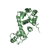
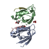
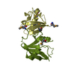
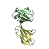
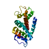
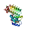
| Title | Three-dimensional structure of triclinic form of de novo design insertion domain, Northeast Structural Genomics Consortium (NESG) Target OR327 | ||||||
 Components Components | Designed protein OR327 Design Design | ||||||
 Keywords Keywords | DE NOVO PROTEIN / Structural Genomics / PSI-Biology / Protein Structure Initiative / Northeast Structural Genomics Consortium / NESG / domain insertion | ||||||
| Function / homology | Ribosomal protein S10 / Alpha-Beta Plaits / 2-Layer Sandwich / Alpha Beta Function and homology information Function and homology information | ||||||
| Biological species | synthetic construct (others) | ||||||
| Method | X-RAY DIFFRACTION / SYNCHROTRON / MOLECULAR REPLACEMENT / Resolution: 2.492 Å | ||||||
 Authors Authors | Kuzin, A. / Su, M. / Seetharaman, J. / Maglaqui, M. / Xiao, R. / Lee, D. / Gleixner, J. / Baker, D. / Everett, J.K. / Acton, T.B. ...Kuzin, A. / Su, M. / Seetharaman, J. / Maglaqui, M. / Xiao, R. / Lee, D. / Gleixner, J. / Baker, D. / Everett, J.K. / Acton, T.B. / Kornhaber, G. / Montelione, G.T. / Hunt, J.F. / Tong, L. / Northeast Structural Genomics Consortium (NESG) | ||||||
 Citation Citation | Journal: Elife / Year: 2015 Title: Precise assembly of complex beta sheet topologies from de novo designed building blocks. Authors: King, I.C. / Gleixner, J. / Doyle, L. / Kuzin, A. / Hunt, J.F. / Xiao, R. / Montelione, G.T. / Stoddard, B.L. / DiMaio, F. / Baker, D. | ||||||
| History |
|
- Structure visualization
| Structure viewer | Molecule: MolmilJmol/JSmol |
|---|
- Downloads & links
-Download
| PDBx/mmCIF format | 4kyz.cif.gz | 142.8 KB | Display | PDBx/mmCIF format |
|---|---|---|---|---|
| PDB format | pdb4kyz.ent.gz | 114.3 KB | Display | PDB format |
| PDBx/mmJSON format | 4kyz.json.gz | Tree view | PDBx/mmJSON format | |
| Others |  Other downloads Other downloads |
-Validation report
| Arichive directory | https://data.pdbj.org/pub/pdb/validation_reports/ky/4kyzftp://data.pdbj.org/pub/pdb/validation_reports/ky/4kyz | HTTPS FTP |
|---|
-Related structure data
| Related structure data |  4ky3SC  5cw9C S: Starting model for refinement C: citing same article ( |
|---|---|
| Similar structure data | |
| Other databases |


-Links
 PDBj
PDBj
- Assembly
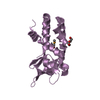
| Deposited unit | 
| ||||||||
|---|---|---|---|---|---|---|---|---|---|
| 1 | 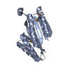
| ||||||||
| 2 | 
| ||||||||
| 3 | 
| ||||||||
| 4 | 
| ||||||||
| Unit cell |
| ||||||||
| Details | monomer,19.75 kD,99.6% |
-Components
| #1: Protein | Design Mass: 19842.973 Da / Num. of mol.: 4 Source method: isolated from a genetically manipulated source Source: (gene. exp.) synthetic construct (others) / Plasmid: pET21_NESG / Production host:  Escherichia coli (E. coli) / Strain (production host): BL21(DE3)+ Magic Escherichia coli (E. coli) / Strain (production host): BL21(DE3)+ Magic#2: Water | ChemComp-HOH / | Water Mass: 18.015 Da / Num. of mol.: 169 / Source method: isolated from a natural source / Formula: H2O Mass: 18.015 Da / Num. of mol.: 169 / Source method: isolated from a natural source / Formula: H2O |
|---|
-Experimental details
-Experiment
| Experiment | Method: X-RAY DIFFRACTION / Number of used crystals: 1 |
|---|
- Sample preparation
| Crystal | Density Matthews: 2.34 Å3/Da / Density % sol: 47.35 % |
|---|---|
| Crystal grow | Temperature: 277 K / Method: microbatch under oil / pH: 8 Details: Protein solution: 100mM NaCl, 5mM DTT, 0.02% NaN3, 10mM Tris-HCl (pH 7.5) . Reservoir solution:0.1M NH4H2PO4 0.1M Tris PEG8K 20% , microbatch under oil, temperature 277K |
-Data collection
| Diffraction | Mean temperature: 100 K |
|---|---|
| Diffraction source | Source: SYNCHROTRON / Site: NSLS  / Beamline: X4C / Wavelength: 0.979 Å / Beamline: X4C / Wavelength: 0.979 Å |
| Detector | Type: MAR CCD 165 mm / Detector: CCD / Date: Apr 12, 2013 |
| Radiation | Monochromator: Si 111 CHANNEL / Protocol: SINGLE WAVELENGTH / Monochromatic (M) / Laue (L): M / Scattering type: x-ray |
| Radiation wavelength | Wavelength: 0.979 Å / Relative weight: 1 |
| Reflection | Resolution: 2.492→30 Å / Num. obs: 23740 / % possible obs: 95.1 % / Observed criterion σ(I): -3 / Redundancy: 3.2 % / Rmerge(I) obs: 0.082 / Net I/σ(I): 15 |
- Processing
| Software |
| |||||||||||||||||||||||||||||||||||||||||||||||||||||||||||||||
|---|---|---|---|---|---|---|---|---|---|---|---|---|---|---|---|---|---|---|---|---|---|---|---|---|---|---|---|---|---|---|---|---|---|---|---|---|---|---|---|---|---|---|---|---|---|---|---|---|---|---|---|---|---|---|---|---|---|---|---|---|---|---|---|---|
| Refinement | Method to determine structure: MOLECULAR REPLACEMENT Starting model: PDB ENTRY 4KY3 Resolution: 2.492→29.457 Å / Occupancy max: 1 / Occupancy min: 1 / FOM work R set: 0.789 / SU ML: 0.33 / Cross valid method: THROUGHOUT / σ(F): 1.52 / Phase error: 28.47 / Stereochemistry target values: ML
| |||||||||||||||||||||||||||||||||||||||||||||||||||||||||||||||
| Solvent computation | Shrinkage radii: 0.9 Å / VDW probe radii: 1.11 Å / Solvent model: FLAT BULK SOLVENT MODEL | |||||||||||||||||||||||||||||||||||||||||||||||||||||||||||||||
| Displacement parameters | Biso max: 69.8 Å2 / Biso mean: 33.308 Å2 / Biso min: 11.46 Å2 | |||||||||||||||||||||||||||||||||||||||||||||||||||||||||||||||
| Refinement step | Cycle: LAST / Resolution: 2.492→29.457 Å
| |||||||||||||||||||||||||||||||||||||||||||||||||||||||||||||||
| Refine LS restraints |
| |||||||||||||||||||||||||||||||||||||||||||||||||||||||||||||||
| LS refinement shell | Refine-ID: X-RAY DIFFRACTION / Total num. of bins used: 8
|
